MOJ
eISSN: 2381-179X


Case Report Volume 5 Issue 2
Department of Pathology, Jawaharlal Nehru Medical College, India
Correspondence: Kafil Akhtar, Professor, Department of Pathology, Jawaharlal Nehru Medical College, Aligarh Muslim University, India
Received: May 02, 2016 | Published: November 25, 2016
Citation: Akhtar K, Saeed N, Ansari HA, et al. Primary carcinosarcoma of ovary with lung metastasis in a young female: a rare case report. MOJ Clin Med Case Rep. 2016;5(2):205-207. DOI: 10.15406/mojcr.2016.05.00129
Primary ovarian carcinosarcomas are rare tumors and account for 1-3% of all ovarian tumors. These tumors mainly affect post-menopausal women, with only 10% incidence in younger women. Ovarian cancer primarily spread to the upper and lower gastrointestinal tract, bladder, liver and spleen. They rarely metastasize to distant sites such as supraclavicular lymph nodes, bone, lung or the brain. The ovarian carcinosarcomas are very aggressive in nature with a poor prognosis. We report a rare case of primary carcinosarcoma of ovary with metastasis to the lung in a 22 year old female with review of literature.
Keywords: carcinosarcoma, lung metastasis, ovary, young female, mesodermal tumor, heterologous tumors, sarcomatous tumors, endometrioid
Primary carcinosarcoma of the ovary is a rare tumor, which accounts for 1-3% of all ovarian malignancies. Histologically both epithelial as well as stromal components are malignant. Carcinosarcoma is also known as malignant mixed mesodermal tumor and further sub classified as “heterologous” or “homologous”, depending on the presence or absence of a stromal mesenchymal component.1 Usually the homologous sarcomatous tumors do not have a better prognosis than the heterologous tumors.2–5 The epithelial serous components are more common than endometrioid and are associated with poor prognosis.2
These tumors mainly affect post-menopausal women, with only 10% incidence in younger women.6 The common clinical complaints are pelvic or abdominal pain, abdominal distention, bowel symptoms and weight loss with associated palpable adnexal mass and ascites. The serum concentration of CA-125 is usually elevated. More than 70% of carcinosarcomas have spread beyond the ovaries at the time of diagnosis.7,8 The spread is primarily intra-abdominal, involving the omentum, upper and lower gastrointestinal tract, bladder, liver and spleen.8 They rarely metastasize to distant sites such as supraclavicular lymph nodes, bone, lung or the brain.9 The primary treatment of ovarian carcinosarcoma is wide surgical excision, followed by chemo-radiation.10 We report a case of primary carcinosarcoma of ovary with metastasis to the lung in a 22year old female with review of literature.
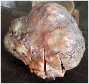
A 22-year-old married female presented in the gynecologic outpatient department with the chief complaints of difficulty in breathing and pain in the lower abdomen since a fortnight. She also complained of fever on and off since 20days with associated weight loss, weakness and fatigue. On physical examination, a mass was felt in the suprapubic region, about 20cm×15cm in size, firm and immobile on palpation. Her laboratory investigations revealed mild microcytic hypochromic anemia and leukocytosis (13,500cells/μl). Liver and kidney functions were normal. Urine analysis revealed no pathology. Pelvic ultrasound and computed tomography revealed a huge and heterogeneous pelvic mass containing solid and cystic areas with ascites measuring 16x13x9cm. Whole body scan revealed no other abnormality. Ascitic tap smear showed only degenerate cells with no viable malignant cells. Chest x-ray revealed left pleural effusion, which on aspiration cytology showed presence of atypical cells suggesting metastatic adenocarcinoma. The serum CA-125 level was markedly raised (126U/ml). Three cycles of neoadjuvant chemotherapy comprising of paclitaxel and carboplatin was administered to relieve her symptoms of breathing, followed by total abdominal hysterectomy with unilateral salpingo-oophorectomy. Laparotomy revealed an unilateral ovarian mass, grossly measuring 13cm×13cm×7cm in size with glistening white and slightly nodular outer surface. Cut surface showed homogenous grayish white solid and cystic areas along with hemorrhage and necrosis (Figure 1). Microscopically section from the tumor showed malignant fascicles of spindle cells with hyperchromatic and pleomorphic nuclei, 30mitosis/10 HPF mixed with areas of necrosis (Figure 2). Necrotic areas might be the carcinomatous component which was responsive to chemotherapy whereas the mesenchymal sarcomatous component was resistant to therapy. Foci of hemorrhage and cystic degeneration were also seen. Histologically, the major part of the tumor consisted of the sarcomatous component which was Van Geison (VG) positive (Figure 3) and showed weak diffuse vimentin positivity on immuno histochemistry (Figure 4). On the basis of clinical findings, gross and microscopic findings, a diagnosis of carcinosarcoma ovary was made.

Figure 1 Grossly the tumor measured 13cm×13cm×7cm in size with glistening white, slightly nodular outer surface with homogenous grayish white solid and cystic cut surface.

Figure 2 Microscopically the section from tumor showed malignant fascicles of spindle cells with hyperchromatic and pleomorphic nuclei, 30 mitosis/10 HPF mixed with areas of necrosis. Haematoxylin & Eosinx40X.
Primary carcinosarcoma of the ovaries is a very rare occurrence, with few available literature.1,2 Pathologically they are biphasic tumors characterized by presence of both malignant epithelial and stromal components.3,4 Recent studies point towards a monoclonal theory of histogenesis for ovarian carcinosarcomas, whereby the metaplasia of the epithelial component gives rise to the sarcomatous component.1 Carcinosarcomas of ovary tend to occur in postmenopausal women with only 10% in young females and show an aggressive clinical behavior.6,11 Our patient was a 22year old female.
It is challenging to differentiate primary ovarian carcinosarcoma pre- operatively from ovarian surface epithelial tumors due to overlapping clinical and radiological findings.5,6 Tumor markers like CA-125 may or may not be raised in ovarian carcinosarcomas, though in our case, serum CA-125 was markedly elevated. Menon et al also showed raised preoperative levels of CA-125 in ovarian carcinosarcoma.1 Cytological study of ascitic fluid does not always be help to detect the malignant component.8 Our patient presented with a large abdominal lump with pain and difficulty in breathing. The CT findings along with cytology of pleural fluid led to the diagnosis of metastatic malignant ovarian tumor. The definitive diagnosis of primary ovarian carcinosarcoma was made on histopathology of the resected abdominal lump. The epithelial component was completely necrosed due to three cycles of neoadjuvant chemotherapy whereas the sarcomatous component was highlighted and further confirmed by immuno histochemistry.
The epithelial component in ovarian carcinosarcoma may be in the form of serous, endometrioid, clear cell adenocarcinoma or squamous cell carcinoma.12 The homologous sarcomatous component may be a tissue native to the ovary like endometrial stromal sarcoma, fibrosarcoma, and leiomyosarcoma and heterologous tissue not native to ovary like chondrosarcoma, rhabdomyosarcoma and osteosarcoma.13 Our case showed the presence of metastatic epithelial adenomatous component in the pleural fluid along with extensive tumor necrosis and fibrosarcomatous stromal component, which were demonstrated both on microscopy and immunohistochemical evaluation. Women usually present with symptoms similar to other epithelial ovarian cancers, but with aggressive behavior.14 About 75-80% of women are diagnosed at stage III or IV of the disease.13 The adverse prognostic factors include older age at diagnosis (60-70years), late stage of the disease and a poor response to platinum-based chemotherapy.12 Amongst all the factors, stage of the disease is the most important predictor of survival.14,15 CA-125 is also considered a prognostic indicator, with pre-operative serum level beyond 75 U/ml associated with a poor outcome.13 Our patient had serum CA-125 value of 126U/ml. The average survival with carcinosarcoma of the ovary is 2 years, with median disease free survival as 11months as compared to 16months for epithelial ovarian cancer.13,14
Wide extensive surgery is considered to be the main treatment modality in ovarian carcinosarcoma.15 The most effective treatment is optimal debulking of the mass lesion, followed by adjuvant chemotherapy, comprising of paclitaxel and platinum based chemotherapy.16 Chemotherapy with cisplatin alone or in combination with ifosfamide, dacarbazine and taxanes have shown response rates of ~ 20%.10,16
The poor prognosis of ovarian carcinosarcomas emphasizes the need for prospective molecular studies to understand better the histogenesis and to implement new chemotherapeutic regimens for better overall survival. Our case highlights an uncommon presentation of the disease, with histopathological and immunohistochemical features.
None.
The author declares no conflict of interest.

©2016 Akhtar, et al. This is an open access article distributed under the terms of the, which permits unrestricted use, distribution, and build upon your work non-commercially.