MOJ
eISSN: 2381-179X


Case Report Volume 13 Issue 4
1Coordinator of International Visiting Medical Students, FIU, USA
2Interna, Medicina, Fundación Universitaria de Ciencias de la Salud, Colombia
Correspondence: Luisa F Garcia, Interna, Medicina, Fundación Universitaria de Ciencias de la Salud, Colombia
Received: November 01, 2023 | Published: November 22, 2023
Citation: Rey CM, Garcia LF. Necrotizing fasciitis and usefulness of diagnostic images: a case report. MOJ Clin Med Case Rep. 2023;13(4):76-78. DOI: 10.15406/mojcr.2023.13.00443
Necrotizing fasciitis is a potentially lethal disease, which is why it is essential to be clear about its pathology and diagnosis. This paper reports the case of a 54-year-old male patient with a history of hypertension and cerebrovascular accident who presented back pain and warm skin with signs of SIRS at the admission. A CT scan was performed with findings consistent on necrotizing fasciitis, however, the delay in the diagnosis contributed the patient required the start of the ACLS protocol; after 4 blue codes the patient died from cardiopulmonary arrest. This case is described in detail and we review the literature on clinical and imaging findings and their usefulness in the diagnosis of this fatal infection.
Keywords: necrotizing fasciitis, CT scan, MRI, imaging findings
A 54-year-old hypertensive male patient with history of cerebrovascular accident was admitted with a clinical onset of 3 months of evolution consisting of back pain of moderate intensity without radiation, not associated with trauma. The character of symptoms were achy and stabbing, there was exacerbating factors including movement and bending over. Patient denies fever and chills. Vital signs at the admission were BP 145/74 mmHg, HR 86 bpm, RR 18 breaths/min, SO2 96%, temperature 98°F (36.7°C). At physical examination patient had shortness of breath and the skin was warm and dry, there was tenderness of the chest wall with swelling and pain to touch. Back findings were edema and erythema associated to pain with crepitus of lower back to palpation. Patient was using a wheelchair because the pain made him very weak and because of that he didn’t allow full physical examination. He was with moderate distress and ill appearing but alert and oriented to person, place, time and situation, normal speech was observed.
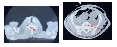
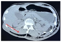
Patient was driven to an abdomen CT (Figure 1) that reported large amount of air in the abdominal wall of uncertain etiology and to a chest CT (Figure 2) that also reported large amount of air in the chest wall. After this, there was performed a CT spine lumbar (lumbar spine CT) with findings of extensive subcutaneous air with thickening of the soft tissues posteriorly for which lead in suspicious diagnosis of necrotizing fasciitis.

Figure 1 Non contrast CT chest scan axial images with no acute infiltrates, no definite mediastinal mass.
There’s large amount of air in the left sided chest wall of uncertain etiology. There’s no fracture of the rib cage.
/p>

Figure 2 Non contrast CT abdomen and pelvis scan axial images with no evidence of pneumoperitoneum or ascitis.
Given the above, invasive ventilation under sedation was started and laboratory test were performed (Table 1), which shown leukocytosis, which, added to signs of hypo perfusion (low BP), it was consider he was in septic shock. It was also noted to be hyperkalemic with initial potassium level of 5 and hyponatremic, as well as severely acidotic with high anion gap metabolic acidosis with a CO2 less than 5. At this time patient experienced a code blue in the emergency department secondary to bradycardia and eventually asystole on monitor. Patient regained return of spontaneous circulation status post ACLS protocol, he was later taken to the ICU and surgery has been consult, however the patient was too week to perform it.
Glucose |
89 mg/dl |
PCO2 |
18.8 mmHg |
PO2 |
235.4 mmHg |
Bicarbonate |
5.1 mEq/L |
Base excess |
-23.4 mEq/L |
FiO2 |
37% |
PT |
25 sec |
INR |
2.28 |
PTT |
29.7 sec |
Lactic acid |
1.2 mmol/L |
WBC |
22.6 x 10 (3) |
RBC |
2.45 x 10 (6) |
HgB |
7.8 g/dl |
Hct |
24.10% |
MCV |
98.4 fl |
MCH |
31.8 pg |
MCHC |
32.4 g/dl |
Platelet count |
501 x 10 (3)/mcL |
Neutrophil abs |
19.86 x 10 (3)/mcL |
Lymphocyte Abs |
0.92 x 10 (3)/mcL |
Monocyte Abs |
1.36 x 10 (3)/mcL |
Eosinophil Abs |
0.02 x 10 (3)/mcL |
Table 1 Laboratories
New laboratory tests were indicated and fluid resuscitation was started with two boluses of NS, also it was established wound care, vasopressin support, anti-thrombotic and antibiotic prophylaxis. Despite all effort, a blue code call was received and unfortunately patient expired after suffering 4 code blues secondary to cardiopulmonary arrest.
Necrotizing fasciitis is a potentially lethal disease with an annual incidence of 1,000 cases annually, and global prevalence of 0.040 cases per 1,000 person-years, which is why it is essential to be clear about its physical pathology and diagnosis.1 Numerous studies show there is a preference for men, with a male to female ratio of 3:1.2 This disease is defined as an infection of the deep soft tissues that causes progressive destruction of the muscle fascia and overlying subcutaneous fat associated with severe signs of sepsis, usually resulting from poly microbial infection.
A direct invasion of subcutaneous tissue is the first step in the pathophysiology of necrotizing soft tissue infections. Most of the time, this is related to processes to external injuries, which allow bacteria from the environment and skin to enter the body. Due to the rapid progression NF carries a median mortality rate of 32.2% and if this entity is not treated with early surgical debridement has a mortality that approaches 100%.3
The organisms (Table 2) related to NF causes tissue damage by releasing exotoxins, which often initiate a complex cascade of immune-related responses including cytotoxic T-cells, cytokine release, and toxic shock syndrome.4 There are two types of NF according to the Society of infectious disease, in both of them microvascular occlusion occurs and may lead to tissue ischemia and subsequent necrosis, the rapid extension of the necrosis constitute barriers to the penetration of antibiotics Type 1 NF is characterized by generating a poly microbial infection;1 commonly its caused by both aerobic and anaerobic bacteria, which is usually related with extensive tissue necrosis and hemodynamic compromise. The complex microbiological profile of offending organisms leads to gaseous infiltration of subcutaneous tissue similar to gas gangrene, due to this, it’s likely to be especially severe in older adults with existing comorbidities (Table 3).4
|
- Clostridium species |
|
- Proteus species |
|
- Escherichia coli |
|
- Bacteroides species |
|
- Enterobacteriaceae species |
Table 2 Common organisms
|
- Diabetes Mellitus, alcoholism |
|
- Peripheral vascular disease |
|
- Malignancy (leukemia, lymphoma) |
|
- Imunicompromise |
|
- Post surgical status |
Table 3 Common comorbidities
Type 2 or mono microbial necrotizing fasciitis can be seen in any patient age group and in tho use with risk factors (Table 4). This kind of infection is associated with gram positive organisms such as group A streptococcus (GAS) and methicillin-resistant staphylococcus aureus (MRSA).1 Endotoxins released by this organisms results in gangrenous myofasciitis and are responsible for some clinical presentations, including toxic shock syndrome. 50% of necrotizing fasciitis occurred by GAS organisms are positive to protein M, which is a virulent factor with anti-phagocytic properties which allows to evade host immune response.
|
- History of blunt trauma |
|
- Penetrating injuries |
|
- Varicella zoster |
|
- IV drug abuse |
|
- Recent surgical procedures |
|
- Burns |
|
- NSAIDs (blunting and delaying of local and systemic immune |
Table 4 Risk factors
During the initial symptoms, NF is characterized by skin erythema progressing beyond margins, edema, warmth, fevers, tenderness, hemorrhagic bullae5 and pain that is disproportionate to the degree of apparent injury. Because this symptoms NF tends to be confused with cellulite. However, as the infection progresses, hemodynamic instability and tissue necrosis can be seen. Organisms that are gas-producing may cause subcutaneous crepitus that can be detected upon palpation of the affected region and skin lesions such as bulla and blisters can occur when the infection is in advanced stages.
Labs usually shows elevate concentrations of creatinine kinase or aspartate amino transferase which also suggest deep tissue infection. Because this pathology presents with non-specific symptoms and is similar to other entities, early clinical suspicion, the use of antibiotics and surgery are key to improving survival.6
Diagnosis
The diagnosis of necrotizing fasciitis is clinical, however, over time, scales such as the LRINEC have been developed, which uses serum levels of the total white blood cell count, hemoglobin, sodium, glucose, creatinine, and C-reactive protein. A score greater than 6 is suggestive of this pathology.
In addition to symptoms and scales, there’s intraoperative findings such as grayish fasciae, loss of resistance of the skin and easy tearing of the deep soft tissues. Histopathologic findings include edematous expansion of inter- and intrafascicular fibrous septa surrounding skeletal muscle bundles, infiltration of inflammatory cells (plasma cells, lymphocytes, neutrophils, and rarely eosinophils), and early fibroblastic proliferation.
Images
The use of images is not required for the diagnosis of this disease, despite this, they are key to reach it. The use of plain radiographs contributes little to the diagnosis due to non-specific findings such as thickening and relative hyperdensity of the soft tissues (rarely visible). However, there is a specific sign which consist in dissecting gas along fascial planes in the absence of trauma7 but is seen in very few patients. Plain radiographs are useful for the detection of soft tissue gas before crepitus is detected, leading to an early diagnosis, which could prevent a fatal outcome.
The ultrasound have big limitations because of the lack of resolution of deeper structures. Findings include an echogenic layer of gas above the deep fascia with posterior dirty acoustic shadowing, hyperechogenicity of the overlying fat, with cobblestone appearance indicating subcutaneous edema, but these findings can also be seen in cellulitis or anasarca,7 that’s why this image is more use to rule out any other underlying patology tan confirming the course of a necrotizing fascitis.
The CT scan is the gold standard image for its evaluation because of the wide availability and high spatial resolution compared to the other images; the most common finding is air in subcutaneous tissue and asymmetric thickening of the fasciae (80%) with adipose- tissue infiltration and usually and soft tissue gas (55%) within fluid collections along subfascial planes.7 The reticular infiltration of the hypodermal fat with the concomitant presence of deep tissue lesions leads into the diagnosis.4 Other characteristics findings are liquefaction necrosis, increases soft-tissue attenuation, inflammatory fat stranding and possible superficial or deep crescenting fluid. The more or less extensive focal inflammation of the hypodermal fat differentiates necrotizing fasciitis from simple stasis edema, which is more symmetric and more diffuse. The sensitivity of CT diagnosing this pathology is 80% but the absence of these findings should not prompt exclusion of the diagnosis because it could be an early disease.
Same as the CT scan, in MRI has a high sensitivity (93%) and the main abnormality is thickening of the deep fasciae, with high signal on T2 images and contrast-enhanced T1 images. T1 images are use for assessing anatomy while T2 images are sequences to look for fascial thickening and edema. Post gadolinium sequences are helpful to delineate the extent of infection, identify abscesses and areas of necrosis.7 The thickening seen is due to fluid accumulation and hyperemia along the necrotic fasciae. Signal intensity from the muscle on T2 images is increased and enhances after contrast injection because of a peripheral superficial distribution. Sensitivity for lesion detection is best with T2 imaging and fat saturation, even compared to post- contrast T1 imaging with fat saturation, which can underestimate the lesions, as a result of necrosis related hypo-perfusion,3 this means the absence of T2 hyperintensity within the deep fascia can essentially exclude a diagnosis necrotizing fasciitis. Besides this, MRI is often not performed because its acquisition is time consuming and will delay treatment.8-10
Concomitant to NF the presence of abscess, gas bubbles as signal-void areas can be seen. Despite of this, it’s important to recognize these are non-specific findings for which the clinic of the patient will always be the key for the differential diagnosis such as cellulitis, radiation-induced damage, osteomyelitis, ruptured popliteal cyst, idiopathic inflammatory myopathies, eosinophilic fasciitis, lymphedema and trauma to the muscles and aponeuroses.
Necrotizing fasciitis continues to be a clinical diagnostic entity (Skin findings, pain out of proportion, and signs of systemic shock), and treatment should not be delayed while waiting for laboratory tests or radiological aids, cause it leads to an almost fatal prognosis. Despite this, in the case of this patient, radiological images were key to its diagnosis. Radiological images are powerful to facilitate early diagnosis, assess the extent of the disease and likewise define surgical planning. The radiological image of choice is CT and MRI because they provide higher sensitivity than other images; the most frequent findings are air in subcutaneous tissue, asymmetric thickening of the fasciae with adipose-tissue infiltration, liquefaction necrosis and inflammatory fat stranding and non-enhancement of the muscular fascia. Finally, it must be taken into account that imaging findings in the early stage may be normal, that means negative studies should not rule out the presence of necrotizing fasciitis, and in the context of high clinical suspicion should not delay the beginning of the treatment.
None.
Authors declare that there is no conflict of interest.

©2023 Rey, et al. This is an open access article distributed under the terms of the, which permits unrestricted use, distribution, and build upon your work non-commercially.