MOJ
eISSN: 2381-179X


Case Report Volume 4 Issue 5
1Addenbrooke's Hospital, United Kingdom
2Lister Hospital, United Kingdom
Correspondence: Phillip Copley, Addenbrooke’s Hospital, Cambridge, United Kingdom, Tel 01223 245151
Received: May 05, 2016 | Published: July 15, 2016
Citation: Copley P, Jaafari FA. Lymphoma as an unusual cause of acute kidney injury: a case report. MOJ Clin Med Case Rep. 2016;4(5):106-107. DOI: 10.15406/mojcr.2016.04.00102
Bilateral hydronephrosis is a rarely encountered condition. We present the case of a 37year old female who presented with urinary frequency, suprapubic pain, fatigue, night sweats, anorexia and weight loss. Ultrasound demonstrated bilateral hydronephrosis and lymphoma was discovered to be the cause on computed tomography guided biopsy. Careful history taking and clinical examination were suggestive of immunosuppression and subsequent HIV testing proved positive. Therapy was commenced simultaneously for both HIV and lymphoma with good clinical response.
Keywords: bilateral hydronephrosis, HIV, lymphoma, acute kidney injury
Hb, haemoglobin; MCV, mean corpuscular volume; CRP, c-reactive protein; CT, computed tomography; FISH, fluorescent in situ hybridization; HAART, highly active anti-retroviral therapy
Lymphoma is a rare cause of bilateral hydronephrosis and subsequent acute kidney injury. Bilateral hydronephrosis can occur in the presence of bladder outlet obstruction or from bilateral obstruction of the ureters. Extramural causes of bilateral obstruction of the ureters include tumours, sarcoidosis and retroperitoneal fibrosis.1
A 37year-old female of West African descent was admitted with urinary frequency and suprapubic pain. Fatigue, night sweats, anorexia and weight loss were also noted on further questioning. She was known to have endometriosis and a previous endometrioma. Her menstrual cycle was regular. She had attended an infertility clinic within the last few months, as she had been unable to conceive over the past six years, and as part of the routine work-up fibroids were noted on pelvic ultrasound. She was on no regular medications, was a non-smoker and family history was unremarkable. On examination, she was apyrexial, haemodynamically stable but tachycardic and had diffuse tenderness of the lower abdomen. A firm mass was noted in the suprapubic region on abdominal examination and renal angle tenderness was elicited. Urinalysis was unremarkable. Initial bloods showed a slight microcytic anaemia (Hb 89g/L; normal=115-165g/L. MCV 74fl; normal=77-95fl), an elevated creatinine at 96umol/L (normal 39-91umol/L) a low albumin (31g/dL; normal=35-50g/dL) and a mildly raised CRP of 22mg/L (normal=<6mg/L). Four months prior to this a creatinine measurement of 59umol/l had been found on routine investigations for infertility. Vaginal swabs demonstrated heavy growth of candida species; tests for Chlamydia trachomatis and Neisseria gonorrhoea proved negative.
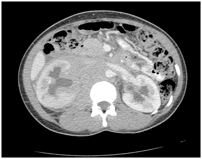
A pelvic mass and bilateral hydronephrosis was noted on ultrasound of the urinary tract. Therefore, a CT scan was arranged. The CT scan (Figure 1) demonstrated a mildly enhancing soft tissue density in the retroperitoneum engulfing the major vessels, ureters and psoas muscles. The mass extended from the diaphragmatic crus to the pelvis. There was bilateral hydronephrosis affecting the right side slightly more than the left. No lymphadenopathy was demonstrable. The features exhibited were consistent with either lymphoma or retroperitoneal fibrosis. In light of her clinical history of recent migration from Africa and her symptoms suggestive of immunosuppression, lymphoma was the more likely in this case. A high-index of suspicion was warranted for infectious causes; however, the weight loss and night sweats indicated lymphoma (B symptoms). The microcytic anaemia, low albumin and slightly elevated C-reactive protein were also suggestive of a chronic condition. Initially bilateral insertion of ureteric double J-stents was performed endoscopically to relieve the ureteric obstruction. This proceeded without complication and post-operatively her creatinine level normalized.

Figure 1 Axial view of CT scan of abdomen showing mildly enhancing soft tissue density in the retroperitoneum causing bilateral hydronephrosis.
A CT-guided biopsy of the mass was performed. Immunohistochemistry, fluorescent in situ hybridization (FISH) studies and molecular analysis demonstrated a high-grade diffuse large B-cell lymphoma. R-CHOP chemotherapy was instigated (Rituximab, cyclophosphamide, doxorubicin, vincristine, and prednisolone).
In light of her presenting symptoms and suspected immunosuppression, tests for HIV and hepatitis were performed. Hepatitis serology was negative. HIV test was positive. She was counseled regarding the new diagnosis of HIV and highly active anti-retroviral therapy (HAART) was commenced. A good clinical response was noted on follow-up with a reduction in viral load and an increase in CD4 count. The lymphoma also responded well to therapy.
Retroperitoneal soft tissue masses may be due to a number of conditions. The differential of a retroperitoneal mass includes primary malignancies and secondary metastatic disease.1 Primary tumours can arise from various structures, and may be benign or malignant. Benign causes include lipomas, adenomas, neurofibromas and cysts.1 Indeed, this patient’s history of leiomy of ibromata raises the suspicion that her disease may have progressed causing encroachment of both ureters. Malignant causes include liposarcomas, neuroblastomas, and lymphomas. Sarcoidosis and granulomatous infections can also mimic this clinical picture, pelvic actinomycosis and tuberculosis being the most commonly cited infectious causes.2–4 Retroperitoneal fibrosis (Ormond’s disease) is characterized by fibrosis of the retroperitoneal tissue that can encase organs in this anatomical location, including the ureters, and has been reported to cause bilateral hydronephrosis.5 It is a rare condition affecting approximately one in a million people per year, with a male predominance (2:1) and commonly affecting those aged 40-60years.6,7 Causes of retroperitoneal fibrosis include malignancies, infections, autoimmune reaction, iatrogenic (surgery, medications and radiotherapy) and idiopathic.1,6,7
In this case it was imperative to initially treat the hydronephrosis in order to protect the kidneys and then identify the cause of obstruction by tissue biopsy. Chemotherapy and immunotherapy are the mainstay treatment in the management of lymphoma. These are not infrequently complicated by neutropenic sepsis and tumor lysis syndrome; thus, patients should be monitored closely during therapy. Lymphoma is a well-recognized complication of an immunosuppressed state and thus it was also important to determine whether there was an underlying cause in this case, especially following her examination findings.8 Indeed, diffuse large B-cell lymphoma is the commonest subtype of lymphoma to occur in HIV patients; this subtype is demonstrated in 80% cases of lymphoma in HIV patients. Prompt identification of her HIV allowed early instigation of HAART therapy to prevent the complications of HIV infection and prevention of further transmission of the virus. This case describes an example of lymphoma in an immunocompromised patient causing bilateral hydronephrosis. It highlights the importance of this differential in acute abdominal pain and kidney injury. It also demonstrates the importance of maintaining a high index of suspicion in a patient with signs of immunosuppression on clinical examination.
None.
The author declares no conflict of interest.

©2016 Copley, et al. This is an open access article distributed under the terms of the, which permits unrestricted use, distribution, and build upon your work non-commercially.