MOJ
eISSN: 2381-179X


Case Report Volume 9 Issue 2
Service de Traumatologie-Orthopédie, CHU Mohammed VI, Oujda, Marooco
Correspondence: Soufiane Aharram, Service de, Traumatologie-Orthopedie, Faculte de Medecine et de, Pharmacie d’Oujda, Marooco
Received: March 09, 2019 | Published: March 27, 2019
Citation: Aharram S, Mounir Y, Benhamou M, et al. Fracture avulsion of the flexor digitorum profundus tendon. case report. MOJ Clin Med Case Rep.2019;9(2):36-37. DOI: 10.15406/mojcr.2019.09.00298
In this report, we present a schematic case of deep flexor tendon avulsion of the fourth finger of the left hand. The lesion consists of an intra-articular fracture of the distal palmar portion of the distal phalanx of the fourth finger, resulting in a large bone fragment, which remains incarcerated at the A4 pulley. The diagnosis was retained preoperatively; reinsetion by "pull out" followed by functional reeducation allowed complete functional restoration after 4 months of trauma.
Keywords : flexor digitorum profundus, fracture, finger jersey, pull out
The traumatic closed fracture of the deep flexor of the fingers is a rare lesion. The first case was described in 1891 by Von Zander.1 In 1960, Boyes et al.,2 and Gunter3 published the first series. This is a common lesion in young athletes. This is usually an intra-articular fracture of the base of the third phalanx of the fingers where the deep flexor tendon of the fingers is inserted. The mechanism is an upset flexion of a finger followed by hyperextension. Careful clinical examination and special radiographic findings make it possible to retain the diagnosis of tearing of the deep flexor tendon of the fingers, which makes it possible to avoid the therapeutic delay and to improve the functional prognosis there after.
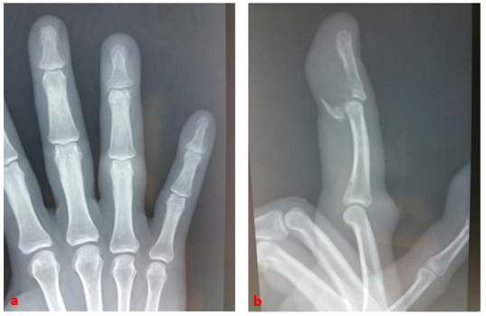
A 49-year-old right-handed police officer suffered a closed trauma to the fourth finger of his left hand as a result of a fall in his height. He then experienced a violent pain concomitant with a cracking sensation, then an impossibility. active flexion of the distal phalanx. The clinical examination, found a finger slightly edematous, ecchymosed. A flexion attitude of the proximal metacarpophalangeal and interphalangeal joints of the same radius, the IPD was in extension and its active flexion impossible (Figure 1). Simple radiographs of the left frontal and lateral annular hands (Figure 2a, b) have led to the diagnosis of anterior marginal articular fracture of the P3 base.

Figure 2a,b Standard radiograph of the front and profile fingers of the annulus confirms fracture tearing of the fragment of the base of the distal phalanx.
The operative indication was posed and the intervention performed under locoregional anesthesia and pneumatic tourniquet, with a palmar zigzag approach according to Bruner; the investigation confirmed the displaced anterior margin fracture of the P3 base, whose reduction and "pull out" fixation enabled the emptiness of the fibrous sheath and distal tendon pulleys (A4, A5) to be demonstrated. FDP (Figure 3).
The bone fragment and tendon were reduced and fixed with an extensible suture attached to a button on the back of the finger by inserting a branch of the suture through the distal fragment (Figure 4). The osteosynthesis was protected by a dorsal immobilization splint kept for four week sallowing passive mobilization by the physiotherapist then the patient began the rehabilitation.
In the patient's final evaluation six months after the procedure, the flexion range of the metacarpophalangeal, proximal and distal interphalangeal joints of the injured finger was 90°, 90° and 70°, respectively. The total active movement of the injured finger was 160°, which is excellent for the Strickland and Glogovac criteria4 (Figure 5).
First described by Boyes et al. in 19602, the Jersey finger translated a distal avulsion of the FDP. The traumatic fractures of the deep flexor of the fingers are rare and little known as evidenced by the small number of series in the literature. The diagnosis must be made urgently by a specialist and the treatmentisal ways surgical. Emergency reintegration results in better functional results: 80% for Mansat and Bonnevialle5, 100% for Tropet et al.6 and Leddy and Packer.7
Regarding the reintegration technique, transosseousre insertion is the mostused and the most reliable. Bunnell's pull-out technique is still widely used, but it tends to be supplanted by micro-brains that have the advantage of their ease of implementation and do not leave externalized material.
If surgical abstention remains the rule in neglected forms and in the sedentary patient, secondary surgical procedures (tendon graft or RPD arthrodesis) may be justified in young motivated manual workers and sportsmen.8
Finger jersey is a pathology of the sportsman, often associated with a fracture of the base of the distal phalanx of a finger. The diagnosis is clinico-radiological, sometimes can go unnoticed.
The evolution is generally favorable if the diagnosis and the therapeutic treatment was early.
None.
The author does not declare any conflict of interest.

©2019 Aharram, et al. This is an open access article distributed under the terms of the, which permits unrestricted use, distribution, and build upon your work non-commercially.