MOJ
eISSN: 2381-179X


Case Report Volume 15 Issue 1
Department of Urology, State University of Campinas - UNICAMP, Brazil
Correspondence: Guilherme Vieira Cavalcante da Silva, Department of Urology, State University of Campinas -UNICAMP, São Paulo, Brazil, Tel +55 19 3521-7481
Received: February 27, 2025 | Published: April 17, 2025
Citation: Silva GVC, Amaro LB, Simões GCS, et al. Forgotten catheter transfixing liver and right kidney: a staged surgical approach. MOJ Clin Med Case Rep. 2025;15(1):9-10. DOI: 10.15406/mojcr.2025.15.00476
A 45-year-old female with recurrent urinary tract infections since adolescence presented with nephrolithiasis, managed by open anatrophic nephrolithotomy in 1997 and percutaneous nephrolithotomy (PCNL) in 2009. In 2018, an MRI incidentally revealed a forgotten catheter transfixing the liver and right kidney, which remained untreated until she experienced worsening of flank pain, hematuria, and pyelonephritis. Upon admission in January 2025, a new CT prompted surgical interventions, including flexible RIRS, PCNL, and laparoscopy for foreign body and calculi removal, with double-J stent placement. This case highlights the importance of proper material counting, imaging control and adequate follow-up to prevent complications.
Keywords: Nephrolithiasis, forgotten catheter, pyelonephritis, urinary tract infections, postoperative imaging
SAH, systemic arterial hypertension; UTI, urinary tract infection; CT, computed tomography; MRI, magnetic resonance imaging; PCNL, percutaneous nephrolithotomy; RIRS, retrograde intrarenal surgery
Retained surgical foreign bodies, including catheters, are rare but significant complications following urological procedures, with an estimated incidence of 0.1-0.2% in abdominal surgeries.1 These foreign bodies can contribute to chronic infections, stone formation, and organ damage, particularly in patients with pre-existing nephrolithiasis. The presence of a retained foreign object may serve as a nidus for stone development, exacerbating conditions like urinary tract infections and pyelonephritis.2 This report describes a complex case involving a 45-year-old female with a forgotten PCNL dilation catheter transfixing the liver and right kidney, underscoring the necessity of intraoperative material counting and diligent postoperative monitoring.
A 45-year-old female with systemic arterial hypertension (SAH) reported recurrent urinary tract infections (UTIs), averaging two episodes per year since adolescence. At age 16 (1997), she was diagnosed with nephrolithiasis in the right kidney and underwent open anatrophic nephrolithotomy. In 2009, she underwent PCNL for stone removal but lost follow-up. In 2018, an abdominal MRI performed for unrelated gastrointestinal symptoms incidentally revealed a catheter in the right kidney, likely retained from previous percutaneous surgery. No immediate intervention was undertaken at that time.
Over the past two years, she experienced worsening right flank pain, initially non-migratory and burning, progressing to colicky pain. She reported gross hematuria (three episodes per month) and was hospitalized for pyelonephritis between 2023 and 2024, requiring prolonged antibiotic therapy. Despite treatment, sporadic colic and hematuria persisted. She was admitted to our service in January 2025. A new abdominal CT confirmed the presence of the forgotten catheter transfixing the liver and entering the right kidney via the superior calyx (Figure 1), along with multiple renal calculi, prompting her hospitalization for surgical intervention (Figure 1).

Figure 1 Preoperative CT image: Right kidney with several calculi and foreign body, which transfixes the liver.
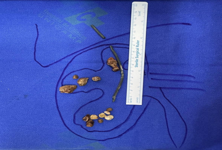
A staged surgical approach was employed. Flexible ureteroscopy and PCNL were performed to remove the distal catheter end and renal stones. The retained foreign body was removed was identified as an 8Fr PTFE dilation catheter from the Amplatz dilation set. Due to pyuria, prolonged surgical time, and bleeding risk, a nephrostomy (Foley catheter) was placed. A subsequent laparoscopic procedure was conducted to extract the hepatic component of the foreign body, followed by anatrophic nephrolithotomy for residual stones (Figure 2– 4). A double-J stent was inserted to ensure ureteral drainage (Figure 2– 4).

Figure 4 Illustration of the forgotten catheter transfixing the liver and entering the right kidney via the superior calyx, with renal calculi in the calyceal system.
Postoperative CT confirmed complete foreign body removal, though some residual calculi remained inaccessible through the procedures performed (Figure 5). The patient was discharged with a Double-J stent for follow-up within one month. Ethics committee approval and informed consent were obtained from the patient (Figure 5).
This case illustrates a rare complication of a forgotten PCNL dilation catheter, likely retained during the 2009 endourological surgery. The catheter’s transfixation through the liver and kidney suggests an intraoperative oversight, contributing to chronic irritation, stone formation, and recurrent infections. The presence of a foreign body acted as a nidus, exacerbating nephrolithiasis and pyelonephritis, with symptom progression due to obstruction and inflammation.2,3
A multi-technique approach was essential. RIRS and PCNL addressed intrarenal pathology, while laparoscopy was necessary for the hepatic and residual intrarenal components. The use of a nephrostomy and double-J stent helped mitigate postoperative risks. The incidental detection via MRI underscores critical need for routine postoperative imaging. The prolonged delay in intervention (2009-2025) likely increased surgical complexity, emphasizing the importance of patient education and adherence to follow-up protocols. Preventive strategies should include meticulous intraoperative material counting and postoperative imaging control after following renal stone treatment procedures.4,5
The successful management of this case via staged surgical interventions highlights the efficacy of a multi-technique approach in removing retained foreign bodies. It also underscores the necessity of postoperative imaging and vigilant follow-up to prevent adverse outcomes. Long-term monitoring remains essential to detect recurrence of nephrolithiasis or infection.4,5
The authors express their gratitude to the staff at the State University of Campinas - UNICAMP for their support during the surgical procedures and patient care.
The authors declare no conflicts of interest.
This research did not receive any specific grant from funding agencies in the public, commercial, or not-for-profit sectors.
CRediT
Wilmar Azal Neto; Writing - Original Draft, Guilherme Vieira Cavalcante da Silva; Writing - Review & Editing, Leonardo Bernardes de Amaro and Gabriel Chahade Sibanto Simões.

©2025 Silva, et al. This is an open access article distributed under the terms of the, which permits unrestricted use, distribution, and build upon your work non-commercially.