MOJ
eISSN: 2381-179X


Case Report Volume 9 Issue 6
1Department of Surgery, Aleppo University Hospital, Syria
2Department of Radiology, Aleppo University Hospital, Syria
3 Faculty of Medicine, University of Aleppo, Syria
Correspondence: Bassel Banjah, Faculty of Medicine, University of Aleppo, Syria
Received: November 13, 2019 | Published: December 3, 2019
Citation: Assaf R, Alsaeed I, otba I, et al. Fetus in fetu: a very rare cause of abdominal mass in a male infant, case report. MOJ Clin Med Case Rep .2019;9(6):150?152. DOI: 10.15406/mojcr.2019.09.00326
Introduction: Fetus in fetu is an extremely rare congenital pathology that has an incidence of one in 500 000 births. This pathology mostly occurs in infants who present with variable manifestations such as a painful abdominal mass and other symptoms caused by the compressive effect of the mass on the host's organs. We report a very rare case that was diagnosed for the first time in Syria.
Case presentation: A 3- month-old male infant was referred to our hospital for abdominal mass associated with hydronephrosis. Abdominal ultrasonography and computed tomography showed a retroperitoneal cystic mass containing multiple calcifications and provoking left hydronephrosis. The diagnosis of teratoma was suspected. Surgical exploration was performed to remove the abdominal mass. The resected mass was then diagnosed as Fetus in fetu due to the clinical and radiological findings after searching about similar reported cases in the medical literature . The patient experienced a good health state after that without recurrence.
Conclusion: Surgeons should keep in their minds this pathology. The diagnosis is made based on a good clinical history and radiological findings. The treatment includes a careful excision of the mass. The patient also should be followed after surgery through clinical examination and radiological investigations.
Keywords: abdominal , ureter, fetu, vertebral, gestation, mass, fetus
Fetus in fetu is an extremely rare congenital pathology that has an incidence of one in 500 000 births.1 The cases reported in the literature are about or fewer than 100 cases in the last two centuries.2 The clinical manifestation is usually resumed to an abdominal mass which is located in the upper right retro-peritoneum space and is often deformed due to compression by the host’s abdominal organ, and there are other manifestations. Our patient presented with abdominal mass and hydronephrosis caused by compression of the ureter via the mass.
A 3 month-old male infant was admitted to our hospital with abdominal mass. He is issued from a non-well controlled pregnancy. The mother had only two ultrasonographic exams in the first trimester of gestation which shew a twin pregnancy and in the third trimester had underwent another one which detected a monofetal pregnancy.
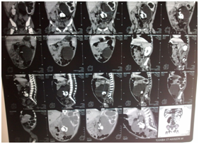
Clinical examination of the patient revealed soft abdominal mass occupying the left hemi-abdomen. Ultrasonography exam had shown a left hydronephrosis associated with polycystic mass measuring 10x15cm containing multiple calcifications. CT scan exam revealed a left hydronephrosis with a thin renal cortex associated with a well-capsulated and heterogeneous mass containing multiple calcifications, measuring 82x66x59mm, localized in the retro-peritoneum space and adherent to the lumbar vertebral column and the left iliac crest, pushing away the aorta and the inferior vena cava to the right side of the abdomen and compressing the left ureter (Figure 1). We suspected with teratoma. Contrast multislice computed tomography of the abdomen was done and it shew a bony mass lying on the left iliac crest and is adherent to the fifth lumbar vertebra and it receives blood perfusion through small vessel arising from the aorta (Figure 2). On surgical laparotomy and exploration using transverse left infra-umbilical incision, we found a large retro-peritoneal cystic mass adherent to the 5th lumbar vertebra and the left iliac crest, pushing the aorta and the inferior vena cava to the right side. There was no adherence with the left kidney but the ureter was compressed causing hydronephrosis.

Figure 1 CT scan shows well-limited cystic mass located in the left lower retroperitoneum containing multiple calcifications provoking a left major hydronephrosis.

Figure 2 Contrast multislice CT scan shows a bony mass attached to the fifth lumbar vertebra( yellow arrow).
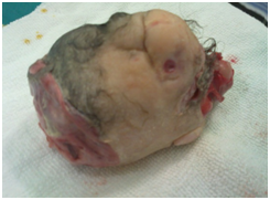
We exteriorized the entire mass carefully. The macroscopic inspection shew a mass weighting 500g covered with fine-hear-bearing-skin, two buds of limbs were seen and a cephalic prominence was noted with primary nose and lips (Figure 3,4). By searching in the literature we found similar cases, so that the diagnosis was made based on the twin gestational history of the mother initially, radiological findings and the macroscopic exam of the mass.

Figure 3 Irregular mass covered with hairy skin with presence of two buds of limbs and a rudimentary face.
Post-operative recovery of the patient was normal and by follow up after three months, he was perfectly well and had no any problems.
Fetus-in-fetu (FIF) is a term first used by Meckel in 18th century and defined by Willis in 1953 as" a mass containing a vertebral axis often associated with other organs or limbs around this axis by which it is differentiated from the highly differentiated teratoma". It is a rare entity where a monozygotic diamniotic and parasitic twin is trapped into its sibling in the early embryonic developmental period and grows inside it via the blood supply of the host circulation .3,4 Although of this definition , there are case reports of FIF with differentiation of tissues without vertebral column.5 Our case was like that, intraoperatively we found an irregular mass covered with hairy skin with two buds of limbs and a rudimentary face, but there was no distinct vertebral axis.
Due to the medical literature, this pathology may be present in newborns, infants, children and adult patients with male superiority. Our patient was a 3 - month-old male infant. Due to our knowledge, this is the first case of Fetus in fetu reported in our country.
Clinical presentation is mostly a painful abdominal mass. However, other localizations were reported as intra-cranial, liver, pelvis, sacrococcygeal region, back, neck, scrotum, mediastinum or oral cavity. In the abdominal localization the mass is frequently localized along the midline retroperitoneally. In our patient the mass was in the lower left retroperitoneum which is a common localization of teratoma.6-9
There are other manifestations secondary to the mass compression of the Fetus in fetu such as hydronephrosis, bowel obstruction, obstructive jaundice, meconium peritonitis, respiratory distress, and vomiting.8 In our case the fetus compressed the left ureter causing hydronephrosis.
Diagnosis before surgery can be suspected on the abdominal plain film, computed tomography and MRI of the abdomen by showing multiple calcifications in the mass which may refer to vertebrae, hip bone, face bones and limbs. Differential diagnosis is teratoma, which is germ cell tumour whithout systemic organization or potential to develop mature tissue.8 It is favorable to do CT angiography before surgical intervention, this can benefit determination blood supply and help in assessing the relationship between the mass and the surrounding anatomic structures.10 In our case, contrast multislice CT of the abdomen was done and it revealed a mass localised above the left iliac crest receiving blood perfusion from small vessels arising from the abdominal aorta (Figure 3).
The recommended treatment is surgical resection of the mass via laparotomy and the specimen should be sent for histopathological examination but in our case we did not send the resected mass for pathological exam because patient's parents rejected that. Although recurrence is uncommon, postoperative clinical, radiological and serological follow up should be done.
Fetus in fetu is a rare pathology that typically presents in infancy. Current imaging modalities may allow us to accurately diagnose the condition before surgery. Surgeons should bear in their mind this rare anomaly in every case with calcificated abdominal mass in children patients. Complete excision is curative and allows confiration of the diagnosis.
None.
None.
None.

©2019 Assaf, et al. This is an open access article distributed under the terms of the, which permits unrestricted use, distribution, and build upon your work non-commercially.