MOJ
eISSN: 2381-179X


Case Report Volume 9 Issue 4
Department of Cardiology, Osmania Medical College/General Hospital, India
Correspondence: Praveen Nagula, Assistant Professor in Cardiology, Department of Cardiology, Osmania Medical College/General Hosptial, India
Received: July 18, 2019 | Published: July 29, 2019
Citation: Chevuru S, Nagula P, Marakkagiri VK, et al. Familial heart hand syndrome - A rare case report. MOJ Clin Med Case Rep. 2019;9(4):84-86 DOI: 10.15406/mojcr.2019.09.00310
Holt-Oram syndrome, the prototype of heart hand syndromes is characterized by abnormalities of the upper limb and congenital cardiac defects. It is rarely seen in 1 in 100,000 children born, with autosomal dominant inheritance. Despite the genetic heterogeneity, it is most frequently caused by a mutation in the TBX5 gene located on chromosome 12. We describe a case of 32years male with morphological abnormalities of upper limbs and atrial septal defect affecting 50% of the siblings by the same disorder.
Keywords Holt-Oram syndrome, hand heart syndrome, autosomal dominant inheritance, atrial septal defect, triphalangeal thumb
Holt-Oram syndrome is a rare heterogeneous genetic disorder with autosomal dominant inheritance, seen in 1 in 100,000 live births.1 Despite genetic heterogeneity, the most frequent mutation is seen in TBX5 gene located on chromosome 12q24.1.2 It is characterized by abnormalities of a radial array and congenital cardiac defects, most commonly atrial septal and ventricular septal defects, hence quoted as atriodigital dysplasia.3 Conduction disturbances are seen in one-third of individuals and may present even in the absence of structural heart disease.4
A 32-year male came with complaints of shortness of breath on exertion of 1-year duration. On physical examination, he had cyanosis, pandigital clubbing, pectus excavatum, and bilateral hand abnormality. He had a triphalangeal thumb and could oppose the thumb with the little finger. Systemic oxygen saturation was 90% at rest and dropped to 82% after exertion. Cardiovascular examination showed displaced apex, a grade 2 left parasternal heave, wide fixed split-second heart sound with a loud pulmonic component.
On detailed history evaluation, he informed to be born out of non-consanguineous marriage with five siblings. His first younger sister died at the age of 16 during the surgery for correction of ASD. Other four surviving siblings were screened. Among them, one female of age 18 was diagnosed to have upper-limb deformity and ostium secundum ASD. The pedigree chart is as below.
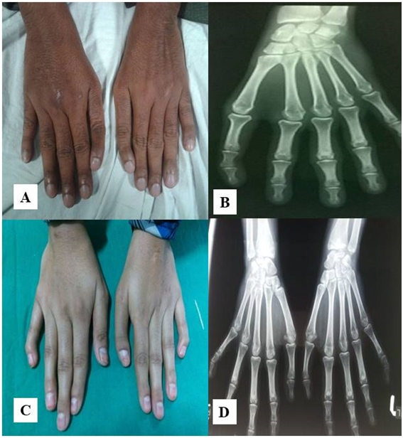
X-ray of his and his sibling's upper limbs showed triphalangeal thumb and carpal bone deformity (Figure 1) (Figure 2) Chest X-ray showed cardiomegaly, enlargement of right heart border and enlarged pulmonary artery. His ECG showed sinus rhythm, right axis deviation, RBBB, first degree AV block.

Figure 2 Photographs of the dorsum of both hands of the patient (A) showing triphalangeal thumb, pandigital clubbing and clinodactyly. (B) The X-ray of the hand of the same patient showing triphalangeal thumb is shown in B. The photo (C) is dorsum of both hands of his sister showing clinodactyly. (D) The x-ray of both hands with wrist bones are shown in photo D.
The 2D-Echo showed ostium secundum atrial septal defect of size 4.4cm with a bidirectional shunt. RA and RV dilated. This was confirmed by TEE. (Figure 3)
2D-echo of his sibling also showed RA and RV enlargement. TEE showed ostium secundum atrial septal defect with left to right shunt. (Figure 4)
The patient was stabilized medically and planned for invasive catheterization study to assess for reversibility of the shunt across the septal defect. Patient’s sibling was planned for ASD closure. Both are under follow up and doing well with medical management.
Group of diseases manifesting with both heart and hand deformities are entitled as Heart Hand Syndromes (HHS), which are rare in occurrence.5 As per the literature, to date heart hand syndromes are of six types. The most common one is the type I also called Holt Oram Syndrome.6 The other syndromes are entitled as Berk Tabatnik Syndrome (Type 2 HHS),Spanish Heart Hand Syndrome (Type3 HHS),Slovenian type (Type 4 HHS), Brachydactyly– long thumb syndrome(Type 5 HHS),Patent Ductus Arteriosus- Bicuspid aortic valve syndrome (Type 6 HHS).7
Familial heart disease with skeletal malformations, first described by Mary Holt and Samuel Oram, was based on four generations of a family with major skeletal manifestation, triphalangeal thumb, and a heart defect, ostium secundum atrial septal defect.8 Here we reported this Holt-Oram syndrome in a family affecting 50% of siblings.
Holt Oram syndrome diagnosis was established by clinical characteristics and if clinical findings are insufficient, a heterogeneous pathogenic variant in TBX5 should be documented, by molecular genetic testing.9
Clinical characteristics are:
Upper limb malformations range from minor radiological abnormalities to phocomelia. Deformities are mainly dysmorphism of carpal bones, aplasia of radius, and shortness of humerus. Abnormal carpal bones present in all affected individuals and identified radiologically may be the only evidence of disease. Mutant gene interferes with embryonic differentiation during the 4th and 5th week of pregnancy when lower limbs are not differentiated. In this case, two siblings had a triphalangeal thumb and carpal bone abnormalities, another sibling might have had a radiological abnormality of carpal bone that was unidentified.
Congenital cardiac defects will be present in 75% of individuals with Holt-Oram syndrome. Almost every type of cardiac anomaly has been reported, either singly or as a part of multiple defects. The most common defect is ostium secundum ASD and VSD located in the muscular trabecular septum. In this case, 3 siblings of a family had ostium secundum ASD, one died during corrective surgery, other developed eisenmenger syndrome secondary to ASD at the age of 32, and the other had ostium secundum ASD.
25% of individuals with Holt-Oram syndrome have conduction abnormalities which can be present even without morphological cardiac defects. Conduction abnormalities range from bradycardia, first-degree AV block to complete heart block.
Most frequently caused by a mutation in the TBX5 gene located on the long arm of chromosome 12. Approximately 80% are due to denovo pathogenic variant. It is inherited as an autosomal dominant disorder. In pregnancies, ultrasound examination may detect congenital heart and upper limb malformations.
In conclusion, we report Holt-Oram syndrome, a rare heart hand syndrome disorder, which affected 50% of siblings in the family with upper limb malformation and ostium secundum ASD.
None.
None.
None.

©2019 Chevuru, et al. This is an open access article distributed under the terms of the, which permits unrestricted use, distribution, and build upon your work non-commercially.