MOJ
eISSN: 2381-179X


Case Report Volume 6 Issue 3
1Department of Community Health Medicine, Hyogo College of Medicine, Japan
2Division of Neurology, Hyogo College of Medicine College Hospital , Japan
3Department of Nuclear Medicine and PET, Hyogo Medical College Hospital, Japan
4Department of Radiological Technology, Hyogo Medical College Hospital, Japan
Correspondence: Kazuo Abe, Department of Community Health Medicine, Hyogo College of Medicine, Japan , Tel +81-798-45-6598, Fax +81-798-45-6597
Received: March 21, 2017 | Published: March 28, 2017
Citation: Abe K, Fukushima K, Maeda Y, et al. Dynamic dopamine transporter imaging for detecting cortical perfusion patterns in Parkinsonism. MOJ Clin Med Case Rep . 2017;6(3):74-76. DOI: 10.15406/mojcr.2017.06.00163
Dopamine transporter (DAT) imaging, a sensitive method to detect the loss of striatal dopamine transporters, can assist in diagnosing neurodegenerative Parkinsonism (PS). Since the early and accurate diagnosis of movement disorders and dementia is critical to ensure optimal clinical management, information on the regional cerebral flow (CBF) is useful for diagnosing PS that involves cerebral cortex disorders, such as progressive supra nuclear palsy (PSP) and corticobasal degeneration (CBD). However, conducting both DAT and CBF imaging is time-consuming and expensive. Thus, we have introduced a novel method for simultaneous DAT and regional CBF imaging.
We recruited eight outpatients with PS and conducted two studies. In the first study, 123I-IMP SPECT imaging was used to measure CBF. In the second study, we performed “dynamic DAT” and conventional DAT imaging. All patients had reduced DAT uptake in the striatum and 123I-IMP SPECT imaging showed four CBF patterns, which correlated well with those observed in dynamic DAT imaging. Since dynamic DAT imaging can be conducted as a series of DAT imaging procedures, it is not only useful for detecting cortical perfusion patterns in PS, but it may also be more time-efficient and cost-effective.
Keywords: dopamine transporter imaging, neurodegenerative parkinsonism, single photon emission computed tomography
DAT, dopamine transporter; DATSANTM, ioflupane123, PS, parkinsonism; PSP, progressive supra nuclear palsy; CBD, corticobasal degeneration, SPECT, single photon emission computed tomography; 123I-IMP, N-isopropyl-p-123I-iodoamphetamine; MMSE, mini mental state examination; FAB, frontal assessment battery; MOCA, montreal cognitive assessment; DLB, dementia of lewy bodies
Dopamine transporter (DAT) imaging using Ioflupane 123I (DATSANTM) is a sensitive method to detect the loss of striatal dopamine transporters1 and can assist in diagnosing neurodegenerative Parkinsonism (PS). Since the early and accurate diagnosis of movement disorders and dementia is critical to ensure optimal clinical management, information on the regional cerebral flow (CBF) is useful for diagnosing PS that involves cerebral cortex disorders, such as progressive supra nuclear palsy (PSP) and corticobasal degeneration (CBD). However, conducting both DAT and CBF imaging is time-consuming and expensive. Thus, we have introduced a novel DAT imaging method that is faster and more cost-effective.
Patients in a Parkinson’s disease clinic located in the Hospital of Hyogo College of Medicine were recruited consecutively. Outpatients were included in the study if they met the UK Parkinson’s Disease Society Brain Bank clinical diagnostic criteria.2 A total of eight patients with PS were recruited. This study was approved by the ethical committee in our institution.
We conducted two studies to measure CBF in patients. Both measurements were carried out in a quiet, dimly lit room with the subjects at rest in the supine position and with their eyes closed.
In the first study, single photon emission computed tomography (SPECT) imaging (Bright View, Philips, Amsterdam, the Netherlands) was used to measure CBF. The resolution was 10.5mm full width half maximum and the computer slice width was 6mm. SPECT data were obtained in a 128×128 format for 64 angles, with 30 s per angle. The SPECT study was initiated 15min after an intravenous injection of 167 MBq of N-isopropyl-p-123I-iodoamphetamine (123I-IMP), and each period of data acquisition was 32min. The filtered back-projection method was used for image reconstruction after preprocessing projection data using the Butterworth filter. A series of slices was reconstructed to be parallel to the orbitomeatal line.
In the second study, surface brain perfusion imaging (which we named “dynamic DAT”) and conventional DAT imaging were initiated straight after and three hours after the intravenous injection of 180 MBq of 123I-N-w-fluoropropyl-2β-carbomethoxy-3β-(4-iodophenyl) nortropane (123I-FP-CIT), respectively. The imaging device and remaining parameters were the same as those used in the first study.
In both studies, data of 123I-IMP SPECT were analyzed using iSSP3 software (Nihon Medi-Physics Co., Ltd.). It is common practice in SPECT analysis to normalize data set to a reference region. In the present work, the algorithm used to normalize each pixel value with stereotactic surface coordinates (x, y, z) to the global perfusion was as follows, Normalized perfusion rate (x, y, z)=Perfusion rate (x, y, z)/Global perfusion rate. The iSSP35_2tZ function in the iSSP3 software was used to compare normal and PS groups in the first and the second study.3
Based on neurological examination and MRI, four patients were diagnosed with PD, one patient with CBD, one patient with PSP, and two patients with dementia of Lewy bodies (DLB).4–6 All patients had reduced DAT uptake in the striatum and 123I-IMP SPECT imaging demonstrated four CBF patterns, which correlated well with the CBF patterns observed in dynamic DAT imaging. Three patients with PD showed normal regional CBF, compared to normal controls. A patient with PD and a patient with PSP showed reduced frontal cortex regional CBF, compared to normal controls. Further, the patient with CBD showed reduced parietoposterior cortex regional CBF compared with those of normal controls. Finally, in comparison to normal controls, the two patients with DLB showed reduced occipital cortex regional CBF.
Results of representative patients
Patient 1
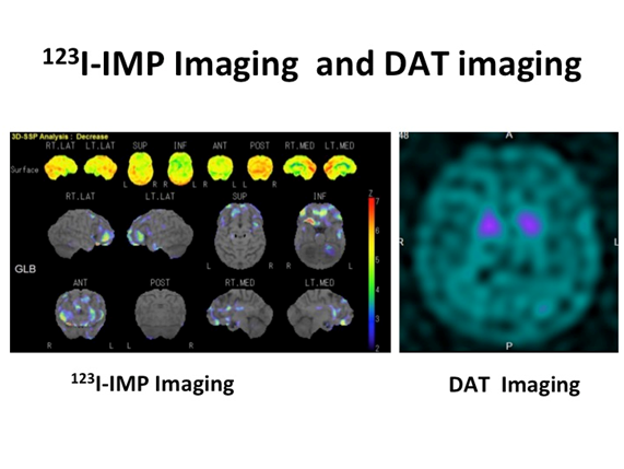
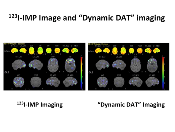
A 66-year-old patient with PD had total scores of 27 out of 30 in the mini mental state examination (MMSE), 12 out of 18 in the frontal assessment battery (FAB), and 24 out of 30 in the Montreal cognitive assessment (MOCA). 123I-IMP SPECT and DAT imaging showed that he had reduced isotope uptake in the frontal lobe and a reduction in striatal dopamine transporters, respectively (Figure 1A). Similar CBF patterns were observed when the 123I-IMP SPECT image was compared to the dynamic DAT image using iSSP3 (Figure 1B).


Figure 1 Patient 1 123I-IMP SPECT and DAT imaging showed that he had reduced isotope uptake in the frontal lobe and a reduction in striatal dopamine transporters, respectively (Figure 1A). Similar CBF patterns were observed when the 123I-IMP SPECT image was compared to the dynamic DAT image using iSSP3 (Figure 1B).
Patient 2
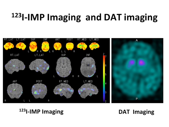
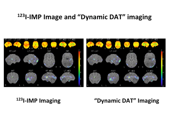
A 69-years-old patient with DLB with visual hallucination scored 25 out of 30, 14 out of 18, and 20 out of 30 in the MMSE, FAB, and MOCA, respectively. 123I-IMP SPECT and DAT imaging showed reduced isotope uptakes in the occipital lobe and a reduction in striatal dopamine transporters, respectively (Figure 2A). The comparison of the 123I-IMP SPECT image with the dynamic DAT image, using iSSP3, demonstrated analogous CBF patterns (Figure 2B).


Figure 2 Patient 2 123I-IMP SPECT and DAT imaging showed reduced isotope uptakes in the occipital lobe and a reduction in striatal dopamine transporters, respectively (Figure 2A). The comparison of the 123I-IMP SPECT image with the dynamic DAT image, using iSSP3, demonstrated analogous CBF patterns (Figure 2B).
The differential diagnosis of PS in a patient is critical to predict the clinical course and ensure optimal clinical management. Until DAT imaging was introduced, clinical signs and symptoms were primarily used for the differential diagnosis of PS. Although DAT imaging has been proven useful for the differential diagnosis of PS, there are increasing cases of PS with dementia, especially DLB,7 where information about CBF is important for precise diagnosis. The differential diagnosis of PS with dementia requires CBF information concerning the cerebral region responsible for cognitive impairment.8 In such a situation, SPECT or positron emission tomography are useful tools for measuring CBF. Minoshima et al.,9 tried to seek antemortem markers to distinguish DLB from Alzheimer's disease (AD). They found that patients with AD and DLB showed significant metabolic reductions involving the parietotemporal association, posterior cingulate, and frontal association cortices. Further, only DLB patients showed significant metabolic reductions in the occipital cortex. They adapted a multivariate analysis and revealed the following three primary components (PC), Patients in PC1 had reduced CBF in the frontal lobe, patients in PC2 had reduced CBF from the frontal lobe through anterior and posterior cingulate gyrus to the posterior lobe, and patients in PC3 had reduced CBF in the posterior lobe. On the basis of these results, they concluded that occipital hypometabolism is a potential antemortem marker that can be used to distinguish DLB from AD. More recently, Thomas et al.,10 conducted a validation study of DAT imaging in the clinical diagnosis of DLB with autopsy as the gold standard. Among patients with DLB, 10% (3 out of 33patients) met the pathological criteria for Lewy body disease but had normal 123I-FP-CIT imaging. This implies that DAT imaging alone is not sufficient to diagnose patients with DLB, with or without mild nigrostriatal dopaminergic pathway pathological changes. Thus, additional CBF imaging may be more helpful for an accurate diagnosis. However, conducting CBF imaging in addition to DAT imaging costs time and money. Thus, since it can be conducted using a series of DAT imaging procedures, the use of dynamic DAT imaging to detect cortical perfusion pattern in PS may be more time-efficient and cost-effective.
None.
The author declares no conflict of interest.

©2017 Abe, et al. This is an open access article distributed under the terms of the, which permits unrestricted use, distribution, and build upon your work non-commercially.