MOJ
eISSN: 2381-179X


Case Report Volume 12 Issue 3
1Department of Pediatric Cardiovascular Surgery, Universidad Nacional Autonoma de México, Mexico
2Department of Pediatric Cardiovascular Anesthesiology, Universidad Nacional Autonoma de México, Mexico
3Department of Pediatric Intensive Care, Universidad Nacional Autonoma de México, Mexico
Correspondence: Carlos Alcantara Noguez, Star Medica Hospital Infantil Privado, Nueva York No 7, Col Nápoles, Ciudad de México, CP 038100, Tel +525559544330
Received: October 07, 2023 | Published: October 26, 2022
Citation: Alcántara C, Godoy G, Pazos E, et al. Double approach in a pediatric patient with dysphagia lusoria due to aberrant right subclavian artery. MOJ Clin Med Case Rep. 2022;12(3):41-42. DOI: 10.15406/mojcr.2022.12.00417
Compression of the esophagus by an aberrant anomalous subclavian artery is usually a finding, and the presence of symptoms is rare. In children, sacrificing said artery can cause hypotrophy of the thoracic limb. We present the case of a 6-year-old female patient with Turner syndrome and dysphagia lusoria, which was resolved with a double approach anastomosing the subclavian artery to the right carotid artery. We consider this approach is safe without compromising the arterial flow of the thoracic member and resolving the digestive symptomatology.
Keywords: dysphagia, lusorian, aberrant subclavian, double approach
An aberrant right subclavian artery (ARSA) is regularly a finding accompanied by congenital heart disease, mainly those involving the aortic arch, however it rarely causes symptoms, in older ages it is a rare but known cause of dysphagia due to its typical retroesophageal course. Treatment of symptomatic cases varies from conservative to surgical.1 Endovascular treatment is not indicated due to mechanical symptoms. Open surgical repair is challenging and requires careful planning.2 In pediatric patients, the sacrifice of the subclavian artery is a common procedure, however, in older ages it is related to hypotrophy of the affected thoracic limb. Simple ligation of the subclavian artery risks “subclavian-vertebral steal” and may be associated with limb length discrepancy without complete resolution of symptoms
A left aortic arch with aberrant right subclavian artery, or lusoria artery (LA), results from regression of the right fourth arch and proximal right dorsal aorta, in combination with persistence of the seventh intersegmental artery arising from the proximal descending aorta. AL has a prevalence of 0.5% to 2% in the general population. LA follows a retroesophageal course in 80% of cases, passes between the esophagus and trachea in 15%, and is anterior to the trachea or main bronchus in 5%. Patients with AL are usually asymptomatic; however, 17% have dysphagia.3
6-year-old female, Turner syndrome, in approach since 3 months for GERD with unsuccessful medical management, endoscopy is performed: presents lax hiatus and images suggestive of GERD; recurrent pneumonia secondary to micro-aspirations, progressive solid and liquid dysphagia. Tomography angiography and 3D reconstruction were performed, where an aberrant right subclavian artery was observed, which arises from the aorta in the terminal portion of the aortic arch, directs its path towards the esophagus, surrounds it and acts as a constrictive ring on the wall. It was decided to perform a two-stage approach: left lateral and right cervical thoracotomy.
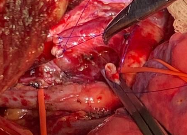
Through left lateral thoracotomy, the aberrant right subclavian artery was identified, which arises directly from the aorta, vascular control is performed, section of the artery and it is carried in the opposite way, dissecting between the adjacent structures, including the wall of the esophagus; In a second cervical stage, an end-to-side anastomosis was performed to the right carotid artery without incident. Oral intake begins at 24 hours without eventualities, and after a follow-up of 3 years, presents no digestive symptoms.
Although the aberrant subclavian artery is of congenital origin, the symptoms associated with illusory dysphagia occur in the fourth and fifth decades of life, the reason being that the vessels are lax in children and young adults. When associated with chronic diseases like hypertension and diabetes, the arteries become stiff.4
Cooley was the first to anastomose the distal subclavian artery to the right common carotid artery.5 The endovascular procedure to treat the stump is safe and minimally invasive. However, there is a risk that these rigid devices within a vessel will erode its wall and cause an esophageal fistula. Multiple reports have documented erosive complications with Amplatzer septal occluders in the heart, even years after placement, with dire consequences. Only long-term follow-up can determine the safety of these devices (Figure 1).6

Figure 1 Section and suture of aberrant subclavian artery, clamped aortic cord to perform suture, and suture reins are left on the distal cord for identification in the cervical approach.
Restoration of anterograde blood flow to the right upper extremity as well as digestive decompression are the goals of surgical treatment. The double approach has been described for the resolution of said pathology, as mentioned by Nelson et al, performing left thoracotomy and median sternotomy, although it could be said that they are more invasive methods, they had a good evolution with discharge in less than 7 days (Figure 2).3
In our experience, a double approach was performed through left thoracotomy, which is frequently performed to resolve pathologies of the aortic arch, pulmonary cercares and patent ductus arteriosus, achieving optimal vascular control, in addition to posterior dissection of the anomalous vessel makes it easy to reproduce. Leaving marking sutures for long, for identification during the second surgical time. A cervical approach, simulating the same approach for peripheral ECMO cannulation, facilitates locating the previously marked cecum ending with an end-to-side anastomosis to the right carotid artery (Figure 3).
We performed control 24 hours later with Doppler ultrasound throughout the arterial vascular path, we started rehabilitation and started aspirin for 3 months.
Currently there is no consensus on what is the best surgical approach to resolve the constrictive effect of the aberrant right subclavian artery? One-stage median sternotomy approaches, two-stage right and left lateral thoracotomy plus a cervical approach have been reported in the literature. There are even descriptions of minimally invasive; each of them clearly offers advantages and disadvantages, studying in detail the anatomy of the aberrant vessel in particular will allow performing the procedure that best suits the patient.
The surgical approach in two stages "Left Lateral Thoracotomy and Cervical" allows to solve dysphagia lusoria in a satisfactory way and without extra complications, in addition to being easily reproducible for cardiovascular surgeons.
The data presented in this article were authorized by the parents of the patient in question for its publication.
None.
Authors declare that there is no conflict of interest.

©2022 Alcántara, et al. This is an open access article distributed under the terms of the, which permits unrestricted use, distribution, and build upon your work non-commercially.