MOJ
eISSN: 2381-179X


Case Report Volume 14 Issue 2
Pulmonology Service, Hospital Clínica San Francisco, Ecuador
Correspondence: Mayra Ortega Ortiz, Pulmonology Service, Hospital Clínica San Francisco, Ecuador Ecuador
Received: February 19, 2024 | Published: June 14, 2024
Citation: Ortiz MO. Clinical case of bone tuberculosis in the foot: involvement of the calcaneus. Diagnostic challenge and possible primary infection. MOJ Clin Med Case Rep. 2024;14(2):52-53. DOI: 10.15406/mojcr.2024.14.00462
Osteoarticular tuberculosis is a rare form of extrapulmonary tuberculosis, and represents a real diagnostic challenge due to its atypical presentation and similarity with other bone pathologies. The case of a 20-year-old male patient with a history of pulmonary tuberculosis treated 5 years ago, who came to the emergency room due to edema, pain and functional difficulty in the ankle and heel, is presented. The computed tomography of the left foot showed a hypodense osteolytic-type lesion that affected two thirds of the calcaneus, thinning of the cortices and involvement in the support area of the calcaneus. Surgical treatment and sample collection were performed, the anatomopathological analysis of which showed the presence of granulomatous cells inside. Tuberculostatic treatment was started, achieving the patient's improvement. The objective is to highlight the importance of early diagnosis based on clinical and radiological findings to facilitate timely intervention and minimize adverse outcomes.
Keywords: tuberculosis, extrapulmonary tuberculosis, osteoarticular tuberculosis, calcaneus
TB, tuberculosis; C, reactive protein PCR; VSR, globular segmentation velocity; AP, anteroposterior
Tuberculosis is a public health challenge caused by Mycobacterium tuberculosis. The vast majority of tuberculosis patients present pulmonary involvement, and only 15% show extrapulmonary manifestations in 2019.1 Osteoarticular tuberculosis is a rare condition that only affects 1-3% of patients with extrapulmonary tuberculosis.2 In Ecuador, the Ministry of Public Health reports that in 2018 it represented 18.46% of total cases of extrapulmonary tuberculosis.3 Tuberculosis of the foot is rare. According to some authors, the talus is the most commonly affected bone, followed by the calcaneus. Clinically, the disease can go unnoticed due to symptoms such as pain, functional limitation and increased volume, which can be associated with multiple pathologies. Diagnosis is often delayed, which can lead to potential complications.1,4,5 This study highlights the different clinical and radiological aspects of the disease to allow early intervention and minimize adverse outcomes. The patient received the corresponding anti-tuberculosis medical treatment according to established protocols, in addition to a surgical intervention.
A 20-year-old male presented with edema, pain and functional difficulty in the ankle and heel of the left foot for 2 months. He went to a private doctor on multiple occasions, who indicated antibiotic treatment and immobilization without clinical improvement, so he went to our service.
Personal history: Pulmonary tuberculosis 5 years ago, which received complete treatment.
Family history: No relevant family history.
Current medication: Not on medication treatment.
Surgical history: No surgical history.
Habits: Does not refer.
Physical exploration: There were no constitutional symptoms of tuberculosis, the left foot with increased volume at the level of the lateral malleolus and heel, pain, decreased ankle mobility in flexion and extension, injury to the external aspect of the ankle.
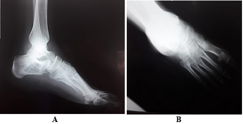
Standard chest x-ray: normal (Figure 1). Anteroposterior (AP) and lateral radiograph of the left ankle. Calcaneus with a delimited circular radiolucent image, with an increase in volume in the soft tissues (Figure 2). Laboratory studies showed positive CRP, increased RSV with lymphocytosis. A vascular Doppler echo of the left lower limb was performed due to the edema, which indicated no vascular compromises. The computed tomography of the left foot shows a hypodense osteological-type lesion, affecting two-thirds of the calcaneus, thinning of the cortices and involvement in the support area of the calcaneus (Figure 3).

Figure 2A Lateral x-ray of left foot shows the calcaneal bone in a delimited circular radiolucent image.
Figure 2B AP x-ray of the left foot shows an increase in volume in the soft tissues.

Figure 3A Coronal section of computed tomography of the left foot shows a hypodense osteological-type lesion.
Figure 3B&3C Sagittal section of computed tomography shows two thirds of the calcaneus, thinning of the cortices and involvement in the support area of the calcaneus.
A sample is taken for culture of common germs which has a negative result. Surgical treatment was decided and a sample was taken and the material was sent to pathological anatomy, which resulted in the presence of granulomatous cells inside. Drainage was performed, bone graft placement was performed before the appearance of bone loss and subsequent immobilization, good postoperative evolutionary control and treatment was started, with isoniazid, rifampicin, ethambutol and pyrazinamide. Tolerance was good, as was the patient's adherence to treatment. The patient evolved favorably, with clinical improvement and progressive decrease in edema and relief of pain, two weeks after starting treatment.
Tuberculosis is a major public health problem, especially in developing countries. Osteoarticular tuberculosis presents an atypical pathogenesis and there is little bibliographic information about it. It can manifest several years after the primary infection, which usually originates in a pulmonary focus and spreads to the bones of the heel or tarsus via blood. These foci remain inactive and are reactivated in situations of immunosuppression, such as malnutrition, childhood, advanced age or chronic diseases. Bone tuberculosis in the foot is more common in young patients or in childhood.1,2 This entity is often confused with many pathologies. The differential diagnosis of these types of lesions, such as chronic osteomyelitis, Paget's disease, sarcomas, and pseudotumor lesions, are usually initially considered as clinical entities, and the diagnosis of tuberculosis is generally ruled out.1–6
In the case presented, the origin of tuberculosis in the calcaneus is primary tuberculosis, possibly due to direct inoculation of the germ that infiltrated the calcaneus. The clinical presentation is unusually subtle, plain radiographs have limited sensitivity and specificity, making sectional imaging techniques such as computed tomography and magnetic resonance imaging more reliable for making an accurate diagnosis.7,8 The treatment in this case was proposed surgically, with the aim of improving the patient's quality of life due to the injuries and another part of the treatment consisted of the administration of anti-tuberculosis drugs in accordance with established standards. The purpose of the presentation of this clinical case is to take into account that, if diagnosed early, this disease has a favorable evolution; Therefore, it is important to consider the clinical characteristics of the patient and the images that provide their main differential diagnoses.9,10
Osteoarticular tuberculosis is a rare entity, with the calcaneus being one of the most commonly affected bones. A multidisciplinary approach, which includes case discussion and evaluation of results, allows for adequate diagnosis, treatment and prognosis in patients with this condition.
None.
The authors declare that there is no conflicts of interest.

©2024 Ortiz. This is an open access article distributed under the terms of the, which permits unrestricted use, distribution, and build upon your work non-commercially.