MOJ
eISSN: 2381-179X


Case Report Volume 13 Issue 2
1Department of Surgery, School of Medicine, Université Catholique du Graben, DRC
2Department of Surgery, Neurosurgery, College of Medicine, Makerere University, College of Health Sciences, Uganda
3Department of Obstetrics and Gynecology, Consolata Hospital Mathari, Kenya
4Department of Internal Medicine, Matanda Hospital, DRC
5Department of Surgery, Université d’AbomeyCalavi, Republic of Benin
6Department of Obstetrics and Gynecology, Mutiri Hospital, DRC
7Department of Obstetrics and Gynecology, Fepsi Hospital, DRC
Correspondence: Olivier Mulisya, Department of Gynecology and Obstetrics, FEPSI Hospital, Butembo, Democratic Republic of Congo, Tel +243997719443
Received: February 24, 2023 | Published: April 10, 2023
Citation: Kamabu LK, Mulisya O, Butala ES, et al. Challenges associated with delayed diagnosis of a giant ovarian mucinous cystadenoma in low resourced settings: a case report. MOJ Clin Med Case Rep. 2023;13(2):25-28. DOI: 10.15406/mojcr.2023.13.00430
Background: The challenge in the diagnosis of giant ovarian cysts (GOC) is a delicate situation for low and middle-income countries where the resources are very limited. GOC have become rare nowadays as they are diagnosed and managed early due to the availability of good imaging modalities. The purpose of this case report is to show how a huge cystic ovarian mass can mislead the diagnosis of multiple pregnancies, ascites, and intestinal tuberculosis in a woman during reproductive age in low-resource countries. The factors associated with the late presentation of giant ovarian cysts in sub-Saharan Africa is also discussed.
Case presentation: The authors present the case of a 36-year-old women, para 4, that was referred to our health center with a grossly distended abdomen wrongly diagnosed as either multiple pregnancy, massive ascites, or intestinal tuberculosis. She was reviewed in different health care facilities without accurate diagnosis. The abdominopelvic ultrasound scan revealed a giant left ovarian cyst. She underwent an exploratory laparotomy where the giant cyst was excised successfully with an uneventful postoperative condition. Histopathology revealed a mucinous cystadenoma.
Conclusion: There are several challenges in accurate and timely diagnosis of GOC in women in reproductive age. Routine abdominal ultrasounds scan use may play a role for guiding to an “early diagnosis“.
Keywords: challenges, giant ovarian mucinous cystadenoma, Sub-Saharan Africa, patient, DRC
β-hCG, beta human chorionic gonadotropin hormone; CA 125, cancer antigen 125; CA 19-9, cancer antigen 19-9; CEA, carcinoembryonic antigen; CT scan, computerized tomography scan; GOC, giant ovarian cyst; LDH, lactate dehydrogenase; MRI, magnetic resonance imaging; USS, ultrasound scan
A giant ovarian cyst (GOC) is any primitive or secondary proliferative process, benign or malignant, of cystic appearance having diameters greater than 10 cm, which growth is not directly linked to hormonal dysfunction. Mucinous cystadenoma of the ovary accounts for about 15% of all ovarian tumors. It is rare and is one of the largest known tumors. It is usually found in women during the reproductive age, rarely before puberty and after menopause.1,2 It is unilateral in 88% spontaneous or iatrogenic causes are more frequent.1 The clinical course can be summarized mainly as an increase in the volume of the abdomen and a sensation of a pelvic mass.2 The diagnosis of GOC is currently facilitated by the use of an abdominopelvic ultrasound scan. The ultrasound scan can be supplemented by an abdominopelvic computerized tomography or by a magnetic resonance imaging. In the case of suspected pelvic neoplasic process, blood sample includes a complete blood count, a quantitative determination of serum beta human chorionic gonadotropin hormone (β-hCG), lactate dehydrogenase (LDH), cancer antigen 125 (CA 125), carcinoembryonic antigen (CEA), and cancer antigen 19-9 (CA19-9). Nowadays, the development of the health system and efficient early detection using advanced technologies has reduced the frequency of diagnosis of these large tumors.3,4 However, they have not entirely disappeared in low developing countries due to poor equipment. Patients usually have poor symptoms which are revealed until the abdomen becomes distended;5,6 this can be misleading during a gynecological examination to suspect pelvic mass in low resource-settings,7 thus, delaying early detection of these tumors until they reach a gigantic size.8 The majority of GOC are mild and are generally treated by surgical excision (cystectomy or salpingo-oophorectomy) or by a percutaneous drainage technique of the cyst, followed by a mini-laparotomy which tends to be the preferred treatment modality for giant ovarian mucinous cystadenomas.2,9-11 Malignant ovarian cysts constitute more than 10% of GOC and are treated by total abdominal hysterectomy with bilateral salpingo-oophorectomy with or without omentectomy.2,4 In the literature, very few cases of GOC mimicking a full-term pregnancy or abdominal ascites or intestinal tuberculosis have been reported. This case would increase the index of suspicion of giant intra-abdominal cysts in women during reproductive age period with abdomino pelvic mass. The factors associated with the late diagnosis of giant ovarian cyst in low resourced settings were also taken into consideration.
Presenting complaint: A 36-year-old woman in reproductive age has been referred to our hospital with a history of abdominal distension, and discomfort for the last 4 years.
History of the presenting complaint: The patient was well until March 2016 when she started to notice a gradual abdominal distension, associated with oligomenorrhea. At this stage, she thought that she was carrying an early pregnancy, and did not seek for antenatal care. After several months, she was worried about the absence of fetal movements, and decided to seek medical care about 36 weeks later. She initially visited a nearby primary healthcare facility where the diagnosis of ascites was made through an abdominopelvic ultrasound scan. She was put on bed rest, diuretics (spironolactone) and fluid restrictions for several days without any improvement. Later, the abdominal discomfort and distension worsened, and was associated with dyspnea. She was then referred to a secondary healthcare facility where a paracentesis was done and about 500ml of straw-colored ascitic fluid was removed to relieve the abdominal symptoms, and was diagnosed of peritoneal tuberculosis based on clinical features. She was then put on anti-tuberculosis treatment with a regimen of rifampicin, isoniazid, ethambutol, pyrazinamide (RHEZ/RE) for almost 6 months, but no bacteriological or advanced laboratories studies were done for confirmation of mycobacterium tuberculosis at this stage. The patient underwent serial ‘paracentesis’ during the first 4 months of anti-tuberculosis treatment, despite all those interventions, the abdominal symptoms (early satiety, bloating, discomfort, constipation) and dyspnea worsened, and had difficulty of performing daily tasks. She was lost to follow up later, and was advised by the relatives to visit other healthcare facilities and traditional medicine for about 1 year without satisfactory outcomes. She was then referred to our tertiary hospital, the Polyclinic of the Adventist University of Lukanga, for further management.
Past medical history: She had no significant medical or surgical history. She had a negative family history of ovarian, uterine, intestine, and breast cancers.
Gyneco-obstetrical history: She is a para 4, with history of 2 spontaneous abortions. She had her menarche at 13 years. Her menstrual cycle was irregular, 4 days in duration and changes the pads 3 times a day with a normal flow. She also reported oligomenorrhea frequently. She was never put on contraception. No prior remarkable gynecological diseases.
Social history: She is a peasant and farmer, does not smoke, neither take alcohol; a mother of 4 children living with her husband.
Review of other system: unremarkable.
Physical examination
General condition: The patient was sick looking, afebrile on touch, not pale, not jaundiced, fully conscious. The patient weighed 75 kg;
Vital signs: Blood pressure: 120/90 mmHg, Pulse rate: 82beats per minute, Respiratory rate 21 cycles per minutes, Temperature: 37.1 degrees Celsius, SPO2 96 percent.
Cardio-vascular system: S1 and S2 heart sounds heard with no change nor added sounds.
Respiratory system: Chest moving with respiration. No crepitation nor crackle heard.
Per abdomen examination
The abdomen was grossly distended, and visible collateral venous circulation in caput medusa. The abdominal circumference of the navel was 105 cm (Figure 1). The abdomen was soft and slightly tender. The liver and the spleen were not palpated. There was a shifting dullness.
Per vaginal examination
Normal perineum and external genitalia. Vagina and cervix were normal. The posterior fornix was not tender.
Other systems: were normal.
Further Investigations
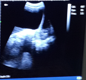
Radiological investigation: Ultrasound was done and revealed a large partitioned anechoic image near the lower pole, centered in a slightly echogenic area, posterior reinforcement (Additional file 1). The size of the image were not easily taken due to the extension of the tumor beyond the monitor screen. . The uterus had no abnormality. The right ovary was visualized and was normal without abnormality. The liver, spleen and renal ultra-sound scans were unremarkable. Intestinal loops pushed back towards the sides, not surrounded by liquid. The abdominopelvic ultrasound concluded that those features were consistent of a giant cyst in favor of the left ovary.

Additional file 1 Trans-abdominal ultrasound image showing a giant left ovary cyst with cumulus oophorus.
Hematological and serological laboratories
Hemoglobin = 12.3g%; Bleeding time = 1min 56 sec; Prothrombine time: 3min 45 sec; HIV serology test: non-reactive.
CA 125: 25 units/ ml,
An undetectable β-hCG.
Diagnosis: Left giant ovarian cyst, probably a mucinous type.
Surgical management
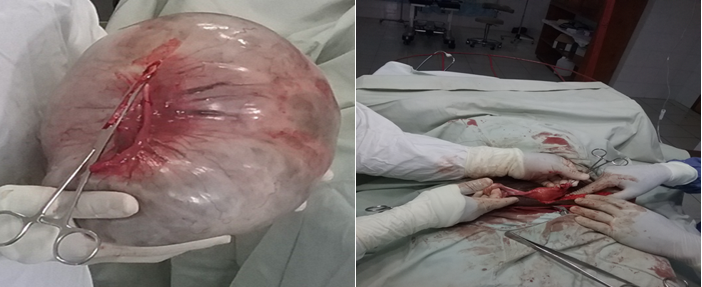
After obtaining a multidisciplinary consultation, a laparotomy under general anesthesia was decided. The patient was operated on with an informed consent. The patient was placed in supine position. The midline incision laparotomy from the xiphoid up to the suprapubic region was performed to allow wide exposure of the large mass. Intraoperatively, there was a large cystic mass. No adhesion was observed. Delicate extirpation of the mass after having removed all the ovarian parenchyma (Figure 2a & b). A left salpingo-oophorectomy and a wedge resection of the right ovary were performed without any hemodynamic and cardiac disturbance (Figure 3a & b), and the cystic mass was resected in total. It measured about 65 × 55 × 34 cm and weighed 24.73 kg (Figure 4a & b). The abdominal wall reconstruction was closed in layers (Figure 5).

Figure 3a & b Total extirpation of the mass and left salpingo-oophorectomy and conservation having removed all the ovarian parenchyma.
Post-operative care: The patient was transferred to the intensive care unit, and had an uneventful postoperative course.
Histology results: The specimen from the left ovary was found to be a benign mucinous cystadenoma characterized by non-hairy columnar cells with nuclei located at the base and apical vacuolation. Examination of the specimens from the right ovary revealed a normal ovarian parenchyma. It was concluded to be benign ovarian mucinous cystadenoma.
Follow-up and outcomes
The wound dressing was uneventful, and she was discharged on the 6th post-operative day with a resolution of her abdominal complaints. She was reviewed in outpatient clinics at 1 and 3 months and by phone call at 6 months.
Giant ovarian cysts have become rare with the development of efficient diagnostic technologies, and early intervention; they definitively require surgical excision.12 Most of the cases are reported in developing regions such as South Asia13 or sub-Saharan Africa.14 The volume described in our patient is the result of an evolution of several years, and a delayed diagnosis as summarized in Figure 6. This delay of getting diagnosis of abdominal mass at an earlier stage of the disease is very frequent in the clinical practice in most of low resources settings due to several reasons.1 In addition to the weakness of the medical referral system, the visit to traditional medicine caused another waste of time of more than a year in the management of this patient. Up to 80% of Africans are still attending traditional medicine for health-related problems according to WHO.2
Giant ovarian cyst is a rare clinical entity. It is usually found in women during the reproductive age like in our case, rarely before puberty and after menopause. Menstrual disorders can be present as in our patient due to the secretion of steroids by the giant cyst; the mechanical complications previously reported by our patient are a function of the volume of the cyst, including dyspnea, early satiety, abdominal discomfort, constipation, and the difficulty in performing daily tasks. The abdominal palpation can reveal a renitent mass, must look for the classic subcostal and suprapubic voids (signs absent if ascites, because collection not circumscribed).15 Usually, there is a dullness with higher convexity, Flot. The Fourestier sign has also been described in the literature and consists of a lumbar sound when seated or squatting due to the discharge of the intestines into the lumbar fossa.1
The diagnosis is usually revealed on ultrasound scan and confirmed by CT or MRI, in the presence of an ovarian mass containing fluid secretions like in our case. It is common to misdiagnosis of a giant cyst versus ascites while performing the ultrasound.
In our patient, the common tumor markers were not measured (LDH, CEA, and CA 125) due to the logistical limitations of our district laboratories. Pre-operative assays of these tumor markers could have allowed guiding the diagnosis.
The mucinous cystadenoma of the ovary is usually benign, and its treatment is surgical consisting in the removal of the mass. The gold standard of resection of ovarian mass includes a laparotomy with intact removal of the mass and involved adnexa, a frozen section evaluation, and subsequent resections according to the pathology result. In our case, there were no available frozen section evaluation. Some authors propose the surgical technique of percutaneous drainage of the cyst, followed by a mini-laparotomy which is a precious example of a safe and effective minimally invasive treatment modality for giant ovarian mucinous cystadenomas.11
In conclusion, very large abdomino-pelvic tumors among female patients in reproductive age are not uncommon in developing countries. This is due to several challenges in accurate and timely diagnosis, also often linked to the patients’ socio-cultural factors, and their ability to visit appropriate health care facilities with competent staffs. In the absence of CT scan imaging facilities, and laboratories for tumor markers assays as it is the case in low-resourced setting, a routine abdominal ultrasound scanning could play an important role in an early management of large abdomino-pelvic tumors.
Written informed consent was obtained from the patient for the publication of this case report.
No financial statement to disclose.
LKK contributed to diagnosis, treatment, postoperative follow-up of patient and wrote the original manuscript with LMK, ESB, BMK, MMV, MKM, BM and RM provided critical revision and correction of the manuscript. OM was involved in the literature review and in critically reviewing the article. HML did the supervision of the work. All authors contributed in intellectual content and approved the final manuscript. All authors have read and agreed to the final manuscript.
The authors would like to thank the patient for her voluntary participation and cooperation in this study.
The authors declare no competing interest.

©2023 Kamabu, et al. This is an open access article distributed under the terms of the, which permits unrestricted use, distribution, and build upon your work non-commercially.