MOJ
eISSN: 2381-179X


Case Report Volume 3 Issue 1
Department of Plastic surgery, Ulster Hospital, Dundonald
Correspondence: Dallan P Dargan, Department of Hand and Reconstructive Microsurgery, National University Hospital Singapore, Tower Block Level 11, 1E Kent Ridge Road, 119228, Singapore, Tel 65 81892978
Received: January 01, 1971 | Published: October 24, 2015
Citation: Dargan DP, Price GJ, Lewis HG. Atypical lipomas of the hand and forearm. MOJ Clin Med Case Rep. 2015;3(1):173-174. DOI: 10.15406/mojcr.2015.03.00052
Three lipomas are presented: lipoma arborescens of the long flexor tendons, a forearm lipoma traversing the interosseous membrane and a thenar eminence lipoma with median nerve compression. Pre-operative magnetic resonance imaging defined the extent of lipomatous infiltration in each case. Excellent outcomes were observed after complete excision.
Keywords: lipoma, hand tumour, hand lipomas, lipoma arborescens, synovial lipomatosis
Lipomas which traverse compartments raise concerns of malignancy. Functional impairment may arise due to lesion bulk, nerve compression and tendon adherence. Lipoma arborescens involving the dorsal wrist and extensor tendons of the forearm and hand has been reported recently.1–3 The condition is extremely rare in this location and flexor tendon involvement contrasts with those in the recent literature.
Case 1: Volar lipoma arborescens
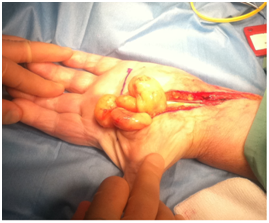
An 81year-old lady presented with gradually enlarging masses in the thenar area, first webspace dorsum and along the flexor tendons of the left hand. Global left hand pain which disturbed sleep was noted, with pincer grip weakness. The medical history included controlled hypothyroidism, hypertension and hypercholesterolaemia. MRI demonstrated a thenar eminence lesion (Figure 1), extending across the flexor tendons in the palm and forearm. Appearances were inconsistent with giant cell tumours or chronic synovitis. High fat content and gadolinium enhancement raised the possibility of liposarcoma. An ultrasound guided biopsy demonstrated benign adiposeand fibrous tissue. The hand was explored under general anaesthetic. Upon opening the carpal tunnel, multiple lipomatous lesions were present along the full extent of the flexor sheath (Figure 2). Each was excised with attached synovial tissue, similar to a rheumatoid synovectomy. A Y-shaped extension of the palmar incision, with division and ligation of the superficial palmar arch, provided access to deeper lesions. Synovial lipomatosis infiltrated the lumbricals and surrounded the flexor tendons. All branches of the median nerve were decompressed. Local anaesthetic was infiltrated and a double lumen Yates drain was placed. Histopathology confirmed benign adipose tissue with fibrovascularstroma and spindle cells. At one month, the wound had healed and finger flexion was full, with resolution of the neuropathy and no recurrence at eight months.

Figure 1 Coronal, sagittal and axial MR images of the left hand, showing lipomatous infiltration around the flexor tendons, and thenar mass.

Figure 2 A volar approach extending across the wrist released a lobulated lipoma within the palm, which extended proximally along the flexor tendons.
Case 2: Forearm lipoma traversing interosseous membrane
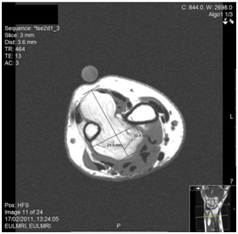
A 60year-old female secretary presented with swelling on the dorsal and volar aspects of her left non-dominant distal forearm and a mild ache at the base of the thumb. The lesion was non-tender and mobile, causing no functional impairment. Past medical history included hypercholesterolaemia, hypertension, gastroesophageal reflux and allergic rhinitis and easy bruising was reported. Ultrasound confirmed a well-defined, hyperechoic lesion, avascular on colour flow imaging, consistent with an intramuscular lipoma.MRI confirmed a 4x2.5x5.5cm lobular fatty tumour of the distal forearm, bridging the anterior and posterior compartments (Figure 3), extending to the distal radioulnar joint, close to the interosseous nerve. Fine-needle aspiration cytology demonstrated benign spindle and epithelioid cells. The possibility of a benign fibrohystiocytic tumour, or low-grade malignancy was raised and synovial sarcoma thought unlikely. Surgical exploration under was performed via a volar approach. A lipomatous lesion deep to the flexor tendons displaced the ulnar artery and nerve. The lesion penetrated the interosseous membrane, which required division for en masse excision. Histopathology reported a benignlipoma. A full functional recovery was noted, no recurrence at two years.

Figure 3 Axial T1 MR Image of the left forearm demonstrating the mass of lipoma crossing the interosseous membrane between radius and ulna.
Case 3: Thenar eminence lipoma with median nerve compression
A 41year-old female classroom assistant with a history of hypertension, hypercholesterolaemia, cigarette smoking and BMI of 33.5 presented with an increasing swelling in her dominant right thenar eminence, compromising thumb movements. Bilateral carpal tunnel symptoms were present since pregnancy 2years earlier. Radiographs confirmed no bony abnormality. MRI confirmed an intramuscular lipoma volar to the trapezium and trapezoid, at the radial border of the carpal tunnel, immediately radial to flexor pollicislongus. The lesion was well defined, with flat signal and fat suppression evident. There was little or no internal architecture, consistent with a lipoma. Surgical excision via a muscle splitting thenar incision was performed under general anaesthetic and tourniquet. A fourth ray incision was used for the carpal tunnel. Histopathological analysis confirmed a benign lipoma, of uniform consistency. The median nerve symptoms resolved, with no recurrence at four years.
Lipomas occur rarely in the hand and fixation to an aponeurosis or confinement to a deep space may restrict mobility of the mass, with absence of the slip sign. Synovial lipomas of the hand may lie within a synovial sheath, or attached to the outer aspect4 and have been described in the palm, wrist and fingers. Lipomas of the midpalmar space may bulge through weak areas of the deep aponeurosis and track along lumbricals.4 Lipoma arborescens differs from lipoma simplex symmetricum due to the ‘tree-like’ pattern of lipomatosis infiltration and is a rare benign disorder.It can occur following scaphoid fracture3 and cause compression neuropathies.5 The expansive branching pattern allows clinical differentiation from angiofibrolipomas which may also involve dorsal wrist tendons.6 Differential diagnosis includes traumatic epithelial cysts, giant cell tumour, liposarcoma and hygroma of tendon sheath (tuberculous tenosynovitis). MRI identifies well-differentiated liposarcoma with sensitivity and negative predictive value quoted as 100%, but specificity of 83% and positive predictive value of 38%.7,8 Malignant transformation of lipomas is exceedingly rare and asymptomatic lipomas can be observed. Surgical dissection around neurovascular structures may be complex9 and recurrence has been noted with incomplete excision.
Atypical lipomas may cause symptoms and concern in this region and MRI and biopsy aid pre-operative planning. Excellent results are possible even in cases requiring complex dissection.
Thanks to Mr P Maginn, Consultant Orthopaedic Surgeon, Musgrave Park Hospital, Belfast and Mr A Brown, Consultant Plastic Surgeon, Ulster Hospital, Dundonald, Northern Ireland for two of the cases discussed.
The author declares no conflict of interest.

©2015 Dargan, et al. This is an open access article distributed under the terms of the, which permits unrestricted use, distribution, and build upon your work non-commercially.