MOJ
eISSN: 2381-179X


Case Report Volume 8 Issue 4
1 Department of stomatology, The 316th Hospital of Chinese People’s Liberation Army, China
2State Key Laboratory of Military Stomatology & National Clinical Research Center for Oral Diseases & Shaanxi Engineering Research Center for Dental Materials and Advanced Manufacture, Department of Anesthesiology, The Fourth Military Medical University, China
3Department of stomatology, Chinese People’s Liberation Army General Hospital, China
Correspondence: Xun Chen, Department of stomatology, The316th Hospital of Chinese People’s Liberation Army, No. A2 Niangniangfu, Xiangshan Road, Haidian District, Beijing, China,
Received: January 01, 1971 | Published: August 27, 2018
Citation: Wang F, Wen J, Du F, et al. A new method to extract mesial impacted teeth: based on proximal and distal diagonal of second molar crown. MOJ Clin Med Case Rep. 2018;8(4):175-177 DOI: 10.15406/mojcr.2018.08.00270
Extraction of mandibular impacted third molars particularly the middle or lower level is the complex alveolar surgery. The conventional procedure often causes many complications. For instance, dry socket, long operation time, severe postoperative pain and slow recovery wound are the common symptoms. Minimal invasive tooth extraction was an important breakthrough to open a new era. This article introduces a new method based on proximal and distal diagonal of second molar crown to extract mesial impacted teeth, which wasn’t reported before.
Keywords: mandibular impacted third molars, teeth extraction
In clinical work, impacted tooth is a common minimally invasive surgery in oral surgery. However, mesial impacted teeth occupy a large proportion. Routine teeth extraction requires hammering, knocking and splitting, which cause large trauma.1 The trend of extraction is to reduce the trauma as much as possible.2 The mandibular impacted third teeth was classified by Winter and he put forward the operative principle of extraction.3 The most common surgery sign is infection because a partially erupted tooth was blocked by the soft tissues and bones.4 The process of the entire surgery includes flap development, bone removal , sectioning, teeth removal and closure.5 However, there is no report to explain how to extract these teeth successfully. Usually we separate the impacted teeth to release the resistance. The general idea to extract mesial impacted tooth is to remove medium resistance crown part by a cutting drill firstly and then we remove the tooth root secondly. However, sometimes we cannot remove the root or residual tooth completely. In regard to the tooth separation guideline, there is no report. Hence, in clinical practice, we summarized this new method of tooth separation to guide the extraction of impacted teeth.
Method
From the clinical practice and summary, we found that proximal and distal diagonal of second molar crown was nearly parallel to the splitting line of the wisdom teeth. Based on this angle, we can divide the teeth to the root bifurcation into two symmetric halves using dental high speed turbine. This novel method can provide a guideline to the surgeons to extract the near medium impacted teeth more conveniently.
In the year 1985, Payne first put forward the concept of “Minimally invasive surgery”.6 The application of minimally invasive surgery can not only elleviate the trauma of patients, but also can promote recovery after surgery. In the routine teeth extraction, chisel, pliers and hammer were the common tools, which would bring fear to patients.7 Hence, minimally invasive concept was becoming increasingly applied in the area of teeth extraction.
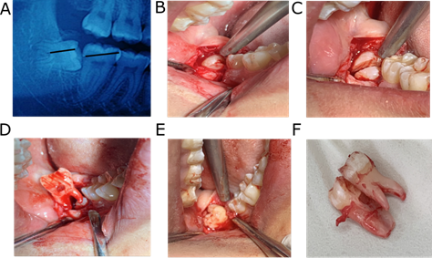
Mandibular impacted third molars, particularly the middle or lower level, is the complex alveolar surgery.8 Firstly, we develop flap, remove bone and split the teeth to release the resistance. In regard to the better teeth separation, there was no report. In our process, we recommend to split the teeth based on the proximal and distal diagonal of second molar crown, which was parallel to the mandibular third teeth separation line. We can successfully divide the teeth to the root bifurcation into two symmetric halves. And then, we remove the teeth’s distal part firstly and the near middle part’s resistance is released completely. This novel method can completely remove the impacted teeth and avoid residual teeth debris. Moreover, to a great extent, this method could reduce the surgical trauma and operation time(F igure 1-3).

Figure 1 Proximal mesial impacted tooth extraction using two-line parallel split tooth method. A) Parallel lines design based on orthopantomography (OPT). B) Flap development and exposure of impacted tooth. C) tooth sectioning following the parallel lines using dental drill. D) Removal of distal part of the tooth. E) Removal of mesial part of the tooth. F) The impacted tooth.

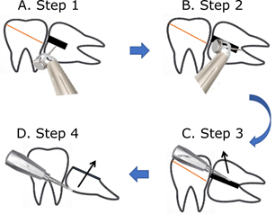
Figure 2 Flow chart for extraction of proximal mesial impacted teeth by the two-line parallel split-tooth method. A) Step 1: The cutting drill is first ground perpendicular to the tooth surface, along the drawn line towards the lingual side, to grind out a groove. B) Step 2: Adjusting the direction of the dental drill in the groove ground in the first step and grinding in the direction of the root bifurcation. C) Step 3: Inserting the dental elevator into the groove and rotating the dental elevator to split the tooth in half and removing the distal part firstly. D) Step 4: Removing the mesial part of the impacted tooth secondly.
This method is a completely novel method to guide the surgeons to successfully split the mesial impacted teeth, which wasn’t report before. In the future, we will conduct a prospective controlled study to further evaluate the advantages of our method compared with the conventional method.
We express our great gratitude to Jianhua Wei for his guidance.
The author declares that they have no conflict of interests.

©2018 Wang, et al. This is an open access article distributed under the terms of the, which permits unrestricted use, distribution, and build upon your work non-commercially.