MOJ
eISSN: 2381-179X


Case Report Volume 6 Issue 3
University Hospital Leuven, Belgium
Correspondence: Sven Bamps, University Hospital Leuven, Belgium
Received: March 25, 2017 | Published: March 30, 2017
Citation: Bamps S, Menten J, Clement P, et al. Selective, bilateral choroid plexus metastases from a renal cell carcinoma presenting as normal pressure hydrocephalus. MOJ Clin Med Case Rep . 2017;6(3):78-79. DOI: 10.15406/mojcr.2017.06.00164
Background: Renal cell carcinoma (RCC) has an unpredictable course and metastases can occur even a long time after nephrectomy. Brain metastases occur in 5 to 10% of RCC and a selective metastasis to the choroid plexus is very rare.
Case description: We report on an 72-year-old male patient with a history of treated RCC who presented after 40months with a classic but severe triad of Hakim and Adams due to hydrocephalus related to a choroid plexus mass in the trigonum of the both lateral ventricles. Because of the severity of symptoms we decided to first treat the symptoms of normal pressure hydrocephalus. Follow-up imaging showed clear disease progression unilateral in the right sided trigonum. Consequently the patient was referred to surgery to remove the right-sided lesion. The left-sided lesion was not resected. The histopathological examination was consisted with a RCC metastasis. The patient was referred for additional systemic treatment.
Conclusion: The frequency of primary tumors causing a choroid plexus metastasis is totally different ad extremely rare. The most common cause was RCC followed by lung, colon and thyroid carcinomas. Surgical resection is an option in case of limited metastasis in patients with RCC. Stereotactic radiosurgery of fractionated external radiotherapy may represent an alternative therapy to achieve local tumor control in the brain. In our case we resected the largest and growing lesion in the right ventricle as we deemed it responsible for the severe symptoms of normal pressure hydrocephalus. The stable left-sided lesion was treated with systemic therapy.
Keywords: renal cell carcinoma, metastasis, choroid plexus
Background
Renal cell carcinoma (RCC) has an unpredictable course and metastases can occur even a long time after nephrectomy. RCC is usually classified as rapidly or slowly progressive and commonly metastasizes to lung (in 70% of cases), lymph nodes (in 60% of cases), liver (in 25% of cases) and bone (in 30% of cases). Brain metastases occur in 5 to 10% of RCC and a selective metastasis to the choroid plexus is very rare with only 10 reported cases in the literature.1–5
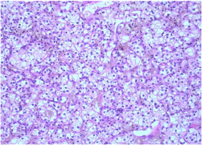
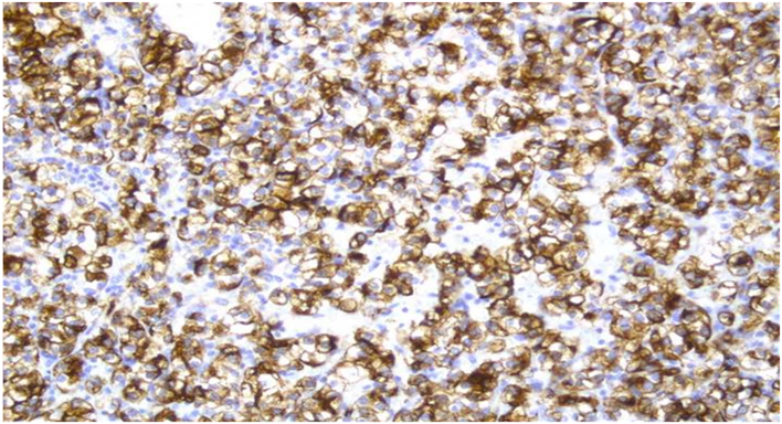
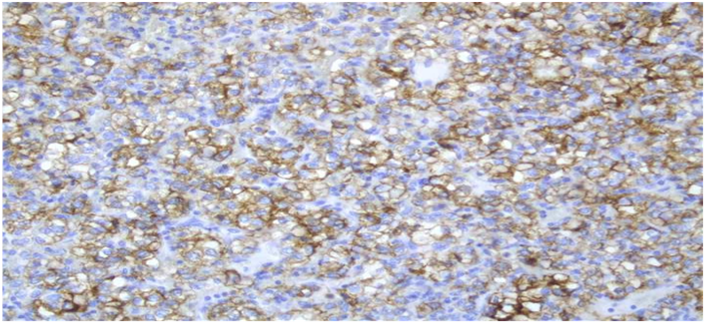
We report on an 72-year-old male patient with a history of treated RCC who presented after 40months with a classic but severe triad of Hakim and Adams (gait disturbance, urinary incontinence and cognitive dysfunction) due to hydrocephalus related to a choroid plexus mass in the trigonum of the both lateral ventricles (Figure 1). Because of the severity of symptoms we decided to first treat the symptoms of normal pressure hydrocephalus initially by external ventricle drainage and finally by VP-shunting to aim for fast symptomatic relieve. Follow-up imaging after 6weeks however showed clear disease progression unilateral in the right sided trigonum (Figure 2). Consequently the patient was referred to surgery to remove the right-sided lesion. The left-sided lesion was not resected. The histopathological examination was consisted with a RCC metastasis as the lesion had the characteristic clear-cell pattern immunoreactive to RCC and CD10 (Figure 3). The patient was referred to the medical oncologist for additional systemic treatment with tyrosine kinase inhibitors.


Figure 3The tumour has the characteristic pattern of a clear-cell renal cell carcinoma with an acinar to trabecular growth pattern of tumour cells with a clear cytoplasm and central nucleus. A delicate fibrovascular network is present between the acinar and trabecular structures (a. H&E stain). The cells are immunoreactive to RCC (B) and CD 10 (C). (Magnification 10x20).
The most common tumors that cause brain parenchyma metastases are lung carcinoma, breast carcinoma and melanoma. The frequency of primary tumors causing a choroid plexus metastasis is totally different ad extremely rare. Raila et al. found only 14cases of choroid plexus metastases from different primary tumors in a 40-year-review. The most common cause was RCC followed by lung, colon and thyroid carcinomas. On MRI-scan, central nervous system metastases from RCC show homogenous contrast enhancement and perilesional edema with characteristic intra- and/or peritumoral flow-voids.1–5 Surgical resection is an option in case of limited metastasis in patients with RCC and was often applied before active systemic treatment options became available. Stereotactic radiosurgery of fractionated external radiotherapy may represent an alternative therapy to achieve local tumor control in the brain. In our case we resected the largest and growing lesion in the right ventricle as we deemed it responsible for the severe symptoms of normal pressure hydrocephalus which is a unique clinical presentation. The stable left-sided lesion was treated with systemic therapy.1–5
None.
The author declares no conflict of interest.

©2017 Bamps, et al. This is an open access article distributed under the terms of the, which permits unrestricted use, distribution, and build upon your work non-commercially.