MOJ
eISSN: 2381-179X


Choanal polyps in nose are frequent cause of nasal obstruction and arise mostly from the lateral wall or the sinuses. We report a case of choanal polyp arising from nasal septum which presented as a mass in the nasopharynx and posed a diagnostic dilemma due to unusual site of attachment. Nasal septum is a very rare site of occurrence and only three cases have been reported in the literature. This was successfully managed with endoscopic nasal surgery. Clinical presentation, differential diagnosis, management, along with review of available literature has also been discussed. Septochoanal polyp should be kept as a differential for mass in the nasopharynx.
Keywords: septochoanal polyp, fibroinflammatory polyp, adult
Polyps are pale prolapsed pedunculated mucosa. These can arise from any mucosal layer of the nasal mucosa.1 A Choanal polyp is defined by their anatomical location and grows towards the choana from a stalk.2 Most of the choanal polyp arises from the ethmoidal infundibulum, and surrounding areas. Choanal polyp arising from the nasal septum or septochoanal polyp are extremely rare and in available literature in English only three such case reports are available.2–5 We report a case of septochoanal polyp in a 25year old man which was arising from the superior aspect of posterior part of nasal septum.
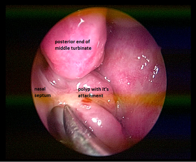
A 25year old man presented with history of progressive bilateral nasal obstruction and snoring for the past two years. There was no history of nasal discharge, post nasal drip, nasal bleeding and his wife reported difficulty in sleeping in the same room with the patient due to snoring. Anterior rhinoscopy was normal. On diagnostic nasal endoscopy a lobulated mass arising from the superior aspect of posterior septum on left side, obstructing whole of the choana was seen (Figure 1). Computed tomography revealed a soft tissue mass occupying whole of the nasopharynx abutting the nasal septum (Figure 2). Paranasal sinuses were normal. Patient was taken up for endoscopic excision and biopsy. Local anesthesia was infiltrated in the pedicle and the stalk of the polyp was resected with the small amount of normal surrounding mucosa. The base of the stalk was cauterized with bipolar cautery. No nasal packing was required and patient was discharged on the same day. Macroscopically the mass was of around 5X2X2cm with lobulated surface and multiple firm nodules were palpable over the surface (Figure 3). On cut section white strands were found, along with few cystic areas (Figure 4). Histopathology revealed a polypoidal tissue mass with marked inflammatory infiltrate containing mostly lymphocytes with lack of Stromal edema and goblet cell hyperplasia; these were suggestive of fibroinflammatory polyp. Postoperative period was uneventful. There was no recurrence in the six months follow up.

Figure 1 Endoscopic photograph on left side showing the polyp filling the posterior choana and its attachment on the superior aspect on posterior septum.

Figure 2 CT scan showing the polyp present in nasopharynx abutting the nasal septum. Sinuses are clear.
Killian described the first choanal polyp in 1906.6 Choanal polyps can arise from maxillary sinus, sphenoid sinus, anterior ethmoids, and middle turbinate.7 Choanal polyps arising from the antrum of the maxillary sinus and are thus termed antrochoanal polyp, from sphenoid sinus as shenochoanal polyp and from ethmoid as ethmochoanal polyp. As nasal septum is covered with mucosa, polyps can arise from it, but these are extremely rare.1,3 Septochoanal polyps are those which arise from the nasal septum and are located in the posterior choana. Only three cases of septochoanal polyps have been reported in the literature.2,4,5 In two of these cases the site of origin was superior portion of posterior nasal septum and in one of the case no site of origin was mentioned. In our case also the site of attachment was superior aspect of posterior part of nasal septum.
Pathogenetically Choanal polyps arise from the recovery process of sinusitis. Obstruction and rupture of mucinous gland leads to expansion of mucinous cyst. Preoperative detection of correct origin of polyp is essential for surgical planning.8 Nasal endoscopy is a noninvasive procedure which can easily detect the origin. CT scan should be done to rule out paranasal sinus involvement and helps to rule out other differrntial diagnosis of a mass in nasopharynx. Septochoanal polyps can be easily excised endoscopically under local anesthesia and a small amount of healthy mucosa surrounding the point of origin of the pedicle should be resected and cuaterised to prevent the recurrence.9 Other benign conditions of nasopharynx such as teratoma, meningoencephalocel, chordoma, paraganglioma, inverted papilloma, adenoid hypertrophy needs to be ruled out.10
None.
The author declares no conflict of interest.

© . This is an open access article distributed under the terms of the, which permits unrestricted use, distribution, and build upon your work non-commercially.