MOJ
eISSN: 2471-139X


Mini Review Volume 5 Issue 4
Department of Veterinary Sciences, University of Chile, Chile
Correspondence: Estefania Flores Pavez, Assistan Professor School of Veterinary Sciences, University of Chile, Santiago, Chile
Received: July 05, 2017 | Published: August 24, 2018
Citation: Mellado BAN, Gino CUF Ricardo OPM, et al. Teeth’s anatomy and dental nomenclatures of small animals. MOJ Anat & Physiol. 2018;5(4):272–275. DOI: 10.15406/mojap.2018.05.00208
Teeth’s anatomy can be described in general, being every tooth formed as a highly calcified structure and having the same sides, and also describes the anatomical particularities of each dental type. Also each species have their dental formula, and regarding this formula, a single tooth can be identified with the usage of a dental nomenclature. There are different dental nomenclatures, such as the anatomical, Triadan (modified), International Federation System and Zsigmondy or of angle; which can be used in the dental record of each patient.
The advances in veterinary odontology have given a great importance to the anatomy of the oral cavity. In this field, teeth are one of the anatomical parts which take place in most of the cases. Knowing the normal structure of each tooth, recognizing a problem in the dental formula, correctly identifying each piece in a written way and being able to transmit this information with the usage of a professional nomenclature are of big importance on a patient’s record. With all the above, the professional can keep a precise record of dental pathologies and lower the risk of committing mistakes in dental procedures such as extractions. The dental nomenclature is the way to identify a dental piece in a fast and precise way.
Teeth are a conical structure situated in opposing rows within the oral cavity.1 They are hard and intensely calcified papillae, white or slightly yellowish, implanted in the jaw’s bone alveoli.2 Teeth are formed by two parts: one contained within the alveolo and other outside it. The junction between both parts is called tecodon.1 each tooth dwells within its own alveolo in the alveolar process of the respective jaw’s bones.3
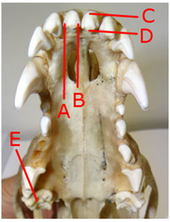
Teeth have five sides (Figure 1):

Figure 1 canine cranium. A) Distal side; B) Mesial side; C) Vestibular side; D) Palatine side (lingual for the inferior jaw); y E) Occlusal side.
The veterinary anatomical nomina6 describe only four sides, since they group the mesial and distal sides, calling them contact side. The dental unit is the sum of the dental piece and its support tissues. The dental piece is formed by a crown, a root or radicle4,7–9 and a neck, which is the crown and root’s (or cement and enamel’s) junction zone,1,7,9 though clinically the terms refers to the portion between the alveolo and the gums.5
The crown is the external and visible part (over the gums), while the root (radicle) is the part immerse in the alveolo, which is not visible in physiological conditions (under the gums).4,9,10 Besides these, the annex tissues are the protective periodontium (marginal gums, adherent gums and alveolar mucosa) and the supportive periodontium (alveolar bone, periodontal ligament and radicular cement).4,10
On the inside of the tooth is found the pulpar cavity which, with the radicular conduct, contain connective tissue, vessels and nerves, besides the dental pulp, that travel through a foramen in the vertex of the root.1,5,10,11 Teeth are innervated by the alveolar nerves, which pass in tunnels through the alveolar process of each bone. It is very difficult to access these nerves. The inferior alveolar nerves are ramifications of the mandibular nerve, giving sensitivity. While on the superior part there are three sets of alveolar nerves: caudal, medium and rostral; branches of the infraorbital nerve.3
Incisives (dentes incisivi)
They are found in the most rostral position of the jaws1 (Figure 2), implanted almost vertically and very closely in the mandibular and maxillary bones. The incisives do not correspond perfectly with their opposing tooth from the other jaw (maxillary against mandibular), but to some portions of them, and can have similar sizes.5,12 They are twelve, divided in four hemi-jaws or quadrants, and are classified in three different types: central (pincers or primaries), intermedium (central or secondary) and lateral (edges or tertiary).9,12 Their labial sides are convex, while their lingual or palatine sides are slightly concave and have a V-shaped crest called the cingula.13
Canines (dentes canini)
They follow the incisives caudally, interrupting the interalveolar or interdentary space (diastema).1 They are large, conical and curve. They fit each other by having the interdentary space in different positions (physiologically the maxillary jaw has its diastema rostrally from the canines, while the mandibular jaw has it caudally from the same). Their roots can measure double the length of their crowns, and oval-shaped, besides having a mesio-distal curvature.4
Premolars (dentes praemolares) and molars (dentes molares)
They are the last elements of the jaw (image 2), conforming the sides of the dentary jaws.1 They have a different size each, being the largest the fourth premolar for the maxillary jaw and the first molar for the mandibular jaw. These teeth also have a special denomination in carnivorous animals, being called carnassial or sectorial teeth.1, 3,5,11 Just as the canines, they do not match the opposing jaw directly, but they fit by their prominences or cones.12
Roots
According to the type of tooth, the number of roots can vary: the incisive, canines and first premolar have one root (Figure 3A); the next premolars have two roots (Figure 3B), except the superior carnasial (fourth premolar) which has three roots; and maxillary molars have three roots (Figure 3C), two vestibular and one lingual, being the vestibular palpable over the maxillary, and the inferior molars have two roots,3,5,9 with the exception of the third lower molar, which has only one root. Whilst cats only have three roots in their superior carnasial tooth.3
Dental formulas
The order of teeth is the same in deciduous (except of not having molars) and permanent dentition. To denominate this order, together with the number of teeth, is that abbreviated methods which describe the number of teeth by quadrant (each jaw is divided in two quadrants), called dental formulas, have been developed.
The dental formula can be expressed in different ways. For example:
2 (I 3/3 C 1/1 P 4/4 M 3/3) = 44.
This formula represents the definitive dentition of the primal plancentary animal.1,8 The “I”s refer to incisives, the “C”s to canines, “P”s to premolars and “M”s to molars. The number shown on the numerator belong to the superior dental jaw, while the denominator to the lower, and the number two before the parenthesis show that this is repeated for both sides of the jaw (for both hemi-jaws) since they are the same in number and characteristics. Capital letters indicate definitive dentition, while low case indicates deciduous dentition. Rosin & Harvey3 use low case for both definitive and deciduous dentitions, but precedes the formula with a “D” for deciduous dentition. A different representation for the dental formula is the one used by Tartaglia & Waugh11
I3, C1, P4, M3
I3, C1, P4, M3
Or the one used by Budras et al.5
|
iii c oppp oo0 |
III C LPPP MM0 |
|
iii c oppp ooo |
III C LPPP MMM |
In this representation deciduous teeth are shown with low case letters, while permanent with capital letters, just like the first case described. Also superior or inferior jaw is the same. The small zeros (“o” in this case) represent a place where there will be a tooth in the definitive dentition, while the big zero (“0”) represent absolute lack of a dental piece in this area for both deciduous and definitive dentition.
Dogs have four premolars on both sides of the maxillary and mandibular bone, as two molars on each side of the maxillary and three on the mandibular. Different from this, cats have less premolars and molars: three and one, respectively, on both sides of the maxillary, and two and one, respectively, on the mandibular. With this, the dental formulas are set like this:
2 (I 3/3 C 1/1 P 3/3) = 28 for puppies,
2 (I 3/3 C 1/1 P 4/4 M 2/3) = 42 for dogs,
2 (I 3/3 C 1/1 P 3/2) = 26 for kittens, and
2 (I 3/3 C 1/1 P 3/2 M 1/1) = 30 for cats.8,11
The difference in the dental formulas of dogs and cats can be due to their diets. Dogs might have more premolars and molars than cats, due to their more omnivorous diet in nature; while cats are strictly carnivorous, being more important to have teeth specialized in tearing and cutting meat.3,11 Brachycephalic animals, because of having a shorter maxillary, lack interdentary space, and also have a more transversally orientation or can even overlap each other, with a possible reduction in the number of teeth of the superior jaw.1
Dental nomenclature
In human as in veterinary odontology, there are nomenclatures to identify each tooth, such as: anatomical, Triadan (modified), International Federation System and Zsigmondy or of angle, among others.
Anatomical: The anatomical nomenclature uses three elements: the letter showing the type of tooth one wants to imply, the number, identifying especifically which of that set is being referred, and the place where this number is written to show the quadrant where it belongs.9 For example, if it is wanted to refer to the fourth definitive premolar of the superior left hemi-jaw, it should be written 4P. While in case of wanting to make reference to the deciduous canine of the inferior right hemi-jaw, it should be written d1.
Triadan (modified): is the adaptation of the nomenclature used in human odotology for veterinary.
In this case each tooth is identified by a three-digit number. The first represents the quadrant where the tooth is, being 1 to 4 definitive dentition, and 5 to 8 deciduous dentition. Number one is the superior right hemi-jaw, and continues clockwise each hemi-jaw (facing the animal) with number two being the superior left hemi-jaw, and so on. The second and third digits are just one number, representing the correlative number of the piece it is referred, without making any reference to the type of tooth it is.
For example, the dental piece number 304 correspond to the definitive canine of the inferior left hemi-jaw, while the piece number 704 is the same piece but deciduous. In this way, the piece 210 corresponds to the second molar of the superior left hemi-jaw. Is important to mention that this dental nomenclature is highly specific for a complete denture and the same number can refer to different pieces depending on the species. A number X08 can be the fourth premolar on a dog or the first molar on a cat, thus it is based on the complete knowledge of the different dental formulas. Besides this, in case of absence of a dental piece, either because of aplasia or loss, the use of this nomenclature can be even more challenging.
International Federation System: This is a system very alike to the last mentioned, with the exception that the first number is separated by a coma from the second one (first digit from the second and third) (Rodríguez, 2002). Using the same example as before, the definitive canine of the inferior left quadrant would be 3,4 (while it was 304 on the other nomenclature). The same risks from the Triadan modified can be identified in this nomenclature.
Zsigmondy or of angles: This nomenclature uses graphic representations (like sides of a cross) referring to the quadrant, followed by a number representing the tooth (exactly as the nomenclatures from before).9 For example, ┌5 correspond to the first premolar of the inferior left quadrant, while ┐6 correspond to the second premolar of the inferior right quadrant or└3 would refer to the third incisive of the superior left quadrant (Figure 4). This nomenclature, besides the numerical problems from the ones before, lack of the differentiation of definitive from deciduous dentition which will be a deficiency while taking notes on a clinical file of each patient.
In veterinary odontology it is of great importance knowing the anatomy of the dental pieces it is worked on. It is of especial importance the number and disposition of roots while performing an extraction, for example, or the side of the tooth while identifying a fracture or dental cavity. To keep a precise and professional record of the procedures a patient have overcome, it is of great importance to be able to identify dental nomenclatures (Figure 5).
With this being said, the anatomical nomenclature would be the one recommended because of its simplicity and completeness. In this case, with just a couple of symbols, all the information needed from a dental piece can be given: which quadrant belongs to, which dentition it belongs to, what type of tooth it is and which specific tooth from its kind.
None.
The authors declare that there is no conflict of interest.

©2018 Mellado, et al. This is an open access article distributed under the terms of the, which permits unrestricted use, distribution, and build upon your work non-commercially.