MOJ
eISSN: 2471-139X


Clinical Report Volume 6 Issue 4
1Head of General surgery Department, Cairo University, Egypt
2General Surgery Department, Cairo University, Egypt
Correspondence: Doaa M Hasan, General Surgery resident at Imbaba general Hospital, El-Giza, Cairo University, Egypt, Tel 02/01062838442
Received: July 31, 2019 | Published: August 14, 2019
Citation: Mounir A, Hasan DM. Schistosomiasis presenting as acute appendicitis with mesenteric nodule filled with bilharzial ova: clinical report. MOJ Anat & Physiol. 2019;6(4):125-126. DOI: 10.15406/mojap.2019.06.00259
Schistosomiasis is a parasitic infestation in humans, commonly in developing countries. The infection manifests itself as a variety of different pathologies, based on the location of the parasite and its eggs. An unusual manifestation is that of a common surgical presentation, acute appendicitis. We present a case of a young male who underwent appendicectomy for acute appendicitis caused by a Schistosomiasis infestation, established upon pathological examination of the resected appendix.
Keywords: mesenteric thrombosis, schistosomiasis infection, diarrhoea, hematuria, normal haemoglobin, ileoceacal junction
Schistosomiasis is a tropical prevalent chronic granulomatous disease that can affect any organ.1 Clinical manifestations of Schistosomiasis vary according to schistosoma species, such as Schistosoma mansoni, Schistosoma haematobium, and Schistosoma japonicum, all of which have similar lifecycles. S. mansoni and S. japonicum cause gastrointestinal symptoms. Without treatment, Schistosoma typically survives in the human body for up to 5 years, but may continue up to 40 years. Chronic infection usually results in life threatening illness due to persistent tissue damage and fibrosis caused by body inflammatory reaction to eggs found in the affected organs. Ordinarily, S. mansoni infects the intestine and liver, while S. haematobium infects the bladder, kidney and ureters. Two uncommon exhibitions of gastrointestinal Schistosomiasis are appendicitis and chronic periodic epigastric discomfort caused by mesenteric thrombosis.2 We document here a case where patient presented with acute appendicitis due to Schistosomiasis infection, established upon histopathological investigation of the excised appendix.
Our case is a 28-year-old male, who was until that time healthy. He came to the emergency department complaining of acute right iliac fossa pain that he had been living through for quite a few days. The pain was not associated with nausea or vomiting, and he had no noteworthy symptoms as headache, hematuria, dysuria, myalgia, arthralgia, cough, diarrhoea or rash.
Proceeding with the clinical examination, the patient had tenderness localized to the right lower quadrant of the abdomen and rebound tenderness. Lab work revealed normal hemoglobin (14.2 g/L) and total leukocyte count (22×109/L), no eosinophilia; platelets were 440×109/L.
The preliminary diagnosis of his case was acute appendicitis.3 Pelvi-abdominal ultrasonography displayed nonspecific intraperitoneal inflammatory change in the area of the terminal ileum and ileoceacal junction, the patient was managed surgically and open appendicectomy was done. Upon surgical excision of the appendix, solitary nodule was found on the mesentery of the ilium about 8 inches from the ileoceacal junction (Figure 1), and excision of the nodule was done.4
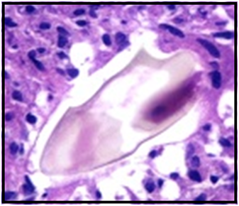
Histopathological analysis of the resected appendix and the nodule exposed acute inflammatory process, marked with copious ova found within the nodule. Further evaluation of the specimen by microbiology and parasitology lab established that these were Schistosoma ova which were designated as ovoid to spherical in shape, and with lateral spines visualized on the eggs (Figure 2). These criteria were indicative of S. mansoni.

Figure 2 S. Mansoni egg inside the granulomatous nodule (H&E [hematoxylin-eosin] stain) 400x magnification.
The patient was referred to the tropical disease hospital for additional assessment and treatment. Later the patient told us that he was born and had lived most of his earliest life in the rural areas of the Delta in Egypt. He was given 60 mg/kg/d of praziquantel divided into 3 doses to which he was well tolerant.5,6

©2019 Mounir, et al. This is an open access article distributed under the terms of the, which permits unrestricted use, distribution, and build upon your work non-commercially.
This is a modal window.