MOJ
eISSN: 2471-139X


Case Report Volume 9 Issue 1
Department of Neurosurgery, Regional Hospital “Dr. Valentín Gómez Farías” Institute of Security and Social Services for State Workers, ISSSTE, Zapopan, México
Correspondence: José Francisco Sánchez Sánchez, Department of Neurosurgery, Regional Hospital “Dr. Valentín Gómez Farías” Institute of Security and Social Services for State Workers, ISSSTE, Av. Soledad Orozco 203, El Capullo, 45100 Zapopan, Jalisco, México
Received: October 27, 2022 | Published: November 2, 2022
Citation: Sánchez-Sánchez JF, Velázquez-Santana H, Serrano-Verduzco JJ. Resection of suprasellar and retrosellar epidermoid cyst by supraorbital approach (minimally invasive approach). illustrative case and literature review. MOJ Anat Physio. 2022;9(1):25-27. DOI: 10.15406/mojap.2022.09.00322
Background: Epidermoid cysts are generally benign congenital lesions that originate from ectodermal tissue; considered as unusual tumors located more frequently in the cerebellopontine angle, being observed in the sellar region in only 10% of cases.
Case description: 56-year-old woman with clinical history of chronic headache and progressive temporal hemianopsia. MRI showed a multilobulated suprasellar and retrosellar lesion that did not enhance the contrast agent, however, T2 sequence showed hyperintensity and diffusion restriction, suggesting an epidermoid tumor.
Conclusion: Due to the multiple approaches to the sellar region, a supraorbital approach (minimally invasive) was chosen, being a direct approach with adequate exposure of the lesion and a lower percentage of complications.
Keywords: Epidermoid cyst, Sellar lesion, Supraorbital approach, Minimally invasive approach, Skull base, Keyhole
Epidermoid cysts (EC) are benign lesions caused by development abnormalities; they approximately represent 0.2 – 2% of the brain tumors. Despite being congenital lesions, they are unusual to be found at an early age due to the lack of clinical manifestations that they present, being mostly diagnosed between 20-40 years.1-3 Lepoire and Pertuiset, having 7 cases of epidermoid cysts, proposed a classification according to their pathogenesis and vascular relationship, dividing them into 3 types: retrosellar, suprasellar and intraventricular; relating them to the distribution of the basilar, carotid and choroidal arteries respectively.4 These lesions are commonly located in the cerebellopontine angle (CPA), followed by the middle floor, prepontine area and chiasmatic region. However, suspicion in the diagnosis will depend on the location and extension of the tumor. Cerebellar signs are commonly found due to the mass effect on the CPA, while hormonal compromise is observed in those lesions with invasion of the sellar region.1-4 In our case, we present an epidermoid tumor that invades the suprasellar, retrosellar and interpeduncular cistern region, being an unusual location, it was decided to be resolved through a transciliary supraorbital approach (minimally invasive).
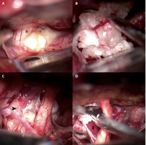
56-year-old female with clinical history of systemic arterial hypertension, carbohydrate intolerance, and dyslipidemia with current management of her comorbidities; no hormonal symptoms or laboratory abnormalities were reported. The patient showed at the Neurosurgery department reporting visual disturbances of her right eye and intermittent oppressive headache in frontal region, being previously assessed by the ophthalmology service and diagnosed with right temporal hemianopsia; however, the imaging study showed a multilobulated cystic lesion in the suprasellar and retrosellar region that invaded the left optochiasmatic cistern with ipsilateral extension that caused retraction of the pituitary stalk. MRI showed restricted diffusion on DWI and increased signal intensity on T2 sequence (Figure 1). For this reason, we decided to perform a left transciliary supraorbital approach; making a 4 cm incision and a craniotomy approximately 30 mm wide x 20 mm high, observing a pearly, soft, and avascular lesion, mostly invading the left optochiasmatic cistern that involved neurovascular structures (left carotid artery, optic nerve left and optic chiasm) (Figure 2). A complete resection of the lesion was performed, without complications, allowing us to discharge the patient in the first 72 hours. The histopathological report showed a keratinized stratified squamous epithelium confirming the diagnosis of an epidermoid tumor (Figure 3).

Figure 1 Preoperative MRI compared with postoperative CT. A. MRI. T2-weighted sagittal view with hyperintense lesion with suprasellar, parasellar and retrosellar extension of left predominance that displaces the pituitary stalk and involves blood vessels. B & D. Hypodense image confined to the sellar region and postsurgical changes that demonstrate absence of occupational lesion with modification of the anatomy towards normality. C. DWI sequence shows a hyperintense lesion with respect to the brain parenchyma, causing diffusion restriction.

Figure 2 Microsurgical resection of a lesion with a cheesy aspect and pearly white coloration. A) Tumor compressing the left optic nerve (✲). B) Removal and limitation of the tumor edge; decompression of the sellar cistern. C) Exposure of carotid bifurcation (➤) and optic chiasm after surgical decompression. D) Tumor resection through the opticocarotid triangle observing the hypophyseal stalk that was previously displaced dorsally (⇞).
Epidermoid cysts are slow growing embryonal tumors caused by aberrant incorporation or incomplete separation of the neuroectoderm during the third to fifth week of gestation with persisting ectodermal tissue during neural tube closure.1,3,5-7 Among other theories, they have been related to the proliferation and metaplasia of adenohypophyseal cells in the sellar region.5,7 Epidermoid tumors account for the 0.2-2% of intracranial tumors and 2.5-7% of CPA lesions; these lesions usually invade extra-axial regions3,7,8 extending through the subarachnoid cisterns and adhering to neurovascular structures, including the perforating arteries.3,9,10
These tumors are generally found in a paramedian or off midline location,10,11 being the CPA the most frequent location (37.3-50%),3,6,11 followed by the middle fossa (15%) and parasellar region (10%).6,11 Vaz-Guimaraes et al. from the Department of Neurosurgery at the University of Pittsburgh School of Medicine, presented a retrospective study of 21 patients with epidermoid and dermoid cysts treated by endoscopic endonasal approaches (EEAs) between January 2005 and June 2014 being one of the largest series of epidermoid cysts of the sellar region counting 8 cases;10 likewise in Somma et al. series, 78 patients were evaluated, presenting rare sellar lesions and showing 4 cases (5%) of epidermoid cysts;12 we can observe that this is an unusual pathology. The clinical presentation varies depending on two important factors: the location of the lesion and its mass effect. Among the most frequent signs and symptoms are: persistent headache (28.6%), dysmetria, ataxia, followed by visual acuity impairment in 32-42.9% (EC of the sellar region), trigeminal neuralgia (14.3%) and hormonal alteration.10,12,13 In those cases in which the epidermoid tumor contains a greater liquid component (cystic) than a solid one, the risk of producing acute symptoms after its rupture increases due to the extravasation of the content (pleocytosis, high protein count and caseous material) into the subarachnoid space, producing meningitis or chemical hypophysitis (10-40%); therefore, draining the cyst in a controlled manner must be considered in order to reduce contact of its content with the parenchyma and the subarachnoid space, avoiding this complication.13,14
Among the imaging studies to consider there is the contrasted computed tomography (CT), which is characterized by a hypodense mass, without contrast enhancement, identifying septations or multilobulated lesions; in the case of sellar lesions we can observe a wide sella turcica, in addition to the density of epidermoid cysts is between −2 and +10 hounsfield units (HU).5,11,14,15 The gold standard is the MRI demonstrating hyperintensity or isointensity of the lesion on T1-weighted sequences due to the cholesterol content of the EC being a solid state that does not enhance on T1-weighted and hyperintense on T2-weighted images. The restricted diffusion on DWI is important to differentiate between EC of dermoid cysts and arachnoid cysts.11,14,15 It is worth mentioning that the risk of malignant transformation of these lesions is rare, we do not have an exact percentage or systematized studies that show the risk factors; in Silva-Vellutini et al. report cases that showed malignant transformation of the epidermoid cyst were complied, observing an increase in the fifth and sixth decade of life, with predisposition in lesions of the CPA, probably because it is the most frequent site of localization;16 showing imaging differences in MR such as hyperintense signal on T1-weighted imaging, as well as perilesional edema and invasion of adjacent structures.15,16 Histopathologic study evidences a keratinizing stratified squamous epithelium, this "flaky" keratin formation is found on a fibrocollagen stroma characteristic of EC.4,13,17
The treatment of these lesions is surgical, there currently some publications that mention the preference of the endonasal endoscopic approach over the open cranial approach, however we must consider two important points previously mentioned; the site of localization and the size of the lesion. During the last decade, the endoscopic endonasal approach has been chosen for sellar, parasellar and infrasellar lesions; however, in Vaz-Guimaraes et al. series they analyzed the benefits of EEA and the factors associated with incomplete resection of the cyst and they exposed the great importance of the remaining tissue; this series, like some others, have concluded that near-total resection (complete resection of the lesion, incomplete of the capsule) or subtotal resection (incomplete resection of the lesion and capsule) lead to a production of keratin that progressively causes the cyst growth, increasing the percentage of recurrence in 27-33.4%.10,14 We must consider that the state of chronic inflammation, secondary to the desquamation of epithelial cells, will allow greater adhesion to vascular and nervous structures causing a greater difficulty in resection;3,9,10 being this reason why total excision is complicated. The EEA has allowed to be an option for the management of tumors of the sella turcica, however, we consider that due to the consistency and characteristics of epidermoid cysts a minimally invasive approach is an excellent option.
In our case, a supraorbital transciliary approach was decided to be used with the objective of a total resection, allowing us to have an adequate working window and manipulation of neurovascular structures due to the tumor´s characteristics and lateral extension; furthermore, decreasing the risk of cerebrospinal fluid (CSF) leak (the most frequent complication in EEA in 33.3%),1,3,10 which, as we mentioned it before, increases the risk of soft tissue infection, abscesses and meningitis.3,6,9,10
The supraorbital approach is an excellent option for pathologies in the anterior and middle floor, including the ventral side of the brainstem; allowing us to limit brain retraction, less tissue manipulation, adequate working window and good cosmetic result;17,18 obtaining greater benefits compared to standard approaches that involve larger diameter craniotomies, larger manipulation of structures and time.
The minimally invasive technique can be complemented with the use of an endoscope to access lesions with lateral extension, cavernous sinus, and contralateral circle of Willis. Among the most frequent complications of the approach are frontal dysesthesia (7.5%), injury to the supraorbital nerve and frontalis palsy (5.5%).17,18 In our case we positioned the patient with a 20º rotation of the head to the right side in order to visualize the suprasellar and retrosellar region, which allowed us an adequate control of neurovascular structures. Despite having displacement of the pituitary stalk secondary to the mass effect, our patient did not developed hormonal alterations as reported in other series such as hyperprolactinemia or hypogonadism secondary to the interruption of prolactin inhibitory factor transport.1,2,6 The prophylactic use of intravenous corticosteroids has benefited the evolution of patients by decreasing the probability of developing chemical meningitis, however, in those cases which suffered cystic rupture or uncontrolled drainage, the risk increases and the use of corticosteroids up to one week after surgery is indicated.11,13
Surgery is the definitive treatment for this type of lesions, taking in count that one of the limitations of total excision is the invasion and adherence of neurovascular structures, which is a challenge for the neurosurgeon. The case we presented is an unusual pathology in a rare growth site, for this reason there isn´t a consensus that establishes the ideal approach for these tumors, we´ve currently observed a greater use of the EEA for the resection of EC of the sellar region; however, a minimally invasive approach offers neurosurgeons an excellent surgical option for a complete resection due to its direct access, less tissue handling and less complications. Due to the previously mentioned data of complications and probability of recurrence of the lesion, the supraorbital transciliary approach allowed us to dissect the neurovascular structures safely to improve the prognosis and survival of our patient.
None.
None.

©2022 Sánchez-Sánchez, et al. This is an open access article distributed under the terms of the, which permits unrestricted use, distribution, and build upon your work non-commercially.