MOJ
eISSN: 2471-139X


Case Report Volume 7 Issue 4
1Conservative Dentistry & Endodontics, Smiline Dental Hospitals, India
2Conservative Dentistry & Endodontics, Navodaya Dental College & Hospital, India
Correspondence: Vijay Venumuddala, Conservative Dentistry & Endodontics, Smiline Dental Hospitals, India, Tel 9000199225
Received: July 29, 2020 | Published: August 14, 2020
Citation: Venumuddala V, Moturi S, Satish, et al. Endodontic management in a rare variant of maxillary third molar with two palatal roots and root canals: a case report. MOJ Anat Physiol. 2020;7(4):121-124. DOI: 10.15406/mojap.2020.07.00301
The maxillary molars, especially the third molars, have the most complicated root canal system in permanent dentition. There are many variations in root canal number system and configuration in maxillary molars. It is imperative and paramount for the clinician to seek out every possible aberration of root canal anatomy for all teeth undergoing treatment. This paper relates a case of a maxillary right third molar with a canal configuration rarely reported in the literature. The tooth had four roots with four root canals, two separate palatal roots (mesiopalatal and distopalatal) with their own distinct canals and orifices. The mesiobuccal and distobuccal roots had normal anatomy. This paper escalates the complexity of maxillary molar variation and is intended to brace clinician’s awareness of the rare morphology of root canal system.
Keywords: distopalatal root, mesiopalatal root, four-rooted maxillary third molar, anatomic variations
A detailed understanding and skill are required to detect and treat the root canal anatomy and its aberrations. The anatomical complexities have been extensively featured in the literature, and the clinician should understand and emphasise the probableaberrations.1,2 Literature review demonstrates an extensive number of variations pertaining to the form, configuration and the number of roots and root canals present in the maxillary molars. Many reports3–5 have mentioned the anatomical variations of the root canal configuration of palatal root of the maxillary molars. The major reason for failure of root canal treatment is due to undetected extra roots or root canals.6 The success of root canal treatment depends on the clinician knowledge about anatomical variations and his/her ability to find and treat the canals.7 Thorough knowledge of root canal anatomy with proper radiographic technique and interpretation are absolute necessity for correct diagnosis and treatment. Preoperative radiographs at different angles helps to interpret extra canals if any and assess root canal anatomy and its aberrations.8 Maxillary and Mandibular third molars usually have wrinkle docclusal anatomy and inadequate access for cleaning and hence are more susceptible for plaque accumulation and decay. Moreover, abnormal eruptive patterns also made them more prone to caries, gingival and periodontal diseases.9 Hence extraction of third molar is usually considered as a routine in dental practice unless demands retention for fixed prosthesis.
Retaining every functional unit of the dental arch which includes third molar is the primary goal of contemporary dental practice.10,11 In some clinical situations third molars are used as abutments for future fixed or removable partial dentures and hence are required to be retained and if root canal therapy is indicated due to existing deep carious lesion involving pulp, extreme care should be taken to thoroughly debride the canals of any inflamed tissue and if left untreated might jeopardize the prosthetic restoration. This case report describes a rare variant of a maxillary third molar with four roots, two buccal roots and two independent palatal roots, each with its own separate canal and its endodontic management.
A 35-year-old Indian male patient had came to the Conservative Dentistry and Endodontics Department complaining of spontaneous pain on the right side of the face from past one week with no significant medical history. On observation clinically, the upper right third molar had a deep carious lesion which was tender on percussion. Electric pulp testing (Analytic Technology, CA) was done and was clinically confirmed as irreversible pulpitis. Patient had missing right maxillary second molar which was extracted few years back and was willing to replace it. Hence, a crown and bridge were planned after the root canal treatment of upper third molar. A pre-treatment radiograph was taken (Figure 1).
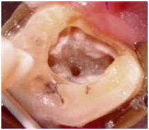
Access cavity preparation was performed under local anaesthesia of 2% lignocaine with 1:2,00,000 epinephrine. Inflamed pulpal tissues located in the pulp chamber were removed and internal anatomy revealed 3 main root canals: mesiobuccal (MB), distobuccal (DB), and palatal (P).Later DG 16 endodontic explorer was used to explore any additional canals. A small haemorrhagic point was noticed around 3-4mm mesial to the main palatal canal indicating a second palatal canal. The regular triangular access cavity shape was modified to a trapezoidal shape in order to gain proper access to the additional canal. The pulp was extirpated, and working lengths of each canal were determined by means of an electronic apex locator (Root ZX; J Morita) and then confirmed by a radiograph (Figure 2). Initial instrumentation of the canals was done with #10 SS files (Dentsply Maillefer).Canal instrumentation and debridement were carried using the crown-down technique with Protaper Rotary files(Dentsply/Maillefer). Apical preparation was performed till F1 for MB, DB canals and F2 for MP and DP canals (Figure 3). Canals irrigated using 2.5% NaOCl and EDTA, access temporized with ZOE cement and appointment was concluded.

Figure 3 Clinical examination showing 4 root canal orifices: 2 buccally and 2 palatally (mirror image).
At the next visit, patient was symptomatic and hence obturation was initiated. Master cone selection was done with the corresponding Pro-taper Gutta-percha cones (Dentsply/Maillefer) (Figure 4) and AH Plus sealer (Dentsply/Maillefer). A radiograph was taken to establish the quality of the obturation (Figure 5). And then the tooth was restored with a posterior composite filling (P60; 3M) and a final radiograph was taken (Figure 6).
Variations in morphology and anatomy, although uncommon can occur in any tooth. The tooth described in this case report is maxillary third molar which had two separate palatal roots, each with a distinct root canal. The majority of endodontic literature describes the maxillary molars as having 3 roots with 3 or 4 root canals.12 The prevalence of 2 palatal roots in maxillary molars is rare and the presence of 2 separate buccal and palatal roots with distinct orifices and foramen is very rare. Whenever indistinct images of palatal roots are presented in preoperative x-ray images taken at different angulations, the clinician should interpret the possibility of two palatal roots and the access cavity is thereby modified accordingly to gain proper instrumentation of the additional canal.13 Shape of access cavity is variable, depending upon the canal configuration, thorough knowledge and properly designed pulp cavities help the clinician to locate and negotiate the root canalanatomy.14 In the present case, the access cavity is made slightly wider palatally in order to accommodate the second palatal canal found mesially thereby converting the conventional triangular access form to trapezoidal (Figure 3). One to three rooted maxillary third molars are more common.15 However, the number of roots/canals in maxillary third molar varies from one to five.16 Sidow et al.10 showed that 15% of maxillary third molars had only one root, 32% had two roots,45% had three roots, where as 7% had four roots. Tomazinho et al.17 reported an unusual case of a maxillary first molar with two palatal roots. Pecora et al.18 showed that 68% had three canals and 34 % had four canals. Stropko19 showed that 60% had three canals, 20% had four canals and 20 % had two canals only.
Anatomical aberrations in permanent maxillary molars are very uncommon, According to Christie et al.20 maxillary molars with two palatal roots are usually found once every 3 years and Peikoff et al.21 speculated that 1.4% of maxillary molars may have two palatal roots. Based on diversion and fusion levels of roots, Christie et al.20 suggested a classification system for four rooted maxillary second molar anomalies. Type I- the two palatal roots are widely divergent and are often long and tortuous. The buccal roots are ‘cow-horn’ shape and less divergent. Type II-all four separate roots are positioned parallel; the roots are short and have blunt apices. Type III-three convergent roots with distinctly divergent fourth distobuccal root. Later Baratto-Filho et al.13 added a Type IV to the original Christie’s classification: the second palatal root is fused with the mesiobuccal root in the coronal two-thirds. The tooth treated in this case appears to be a combination of Type I and Type II variety according to the Christie’s classification-The buccal roots are less divergent and cow horn shape like Type I and palatal roots are short and blunt like Type II but divergent unlike parallel. This makes the present case report even more unique.
A more recent study was conducted by Gu Y et al.22 in Chinese population on 1365 maxillary first and second molars with the help of CBCT imaging and found the incidence rate of 0.07% and 0.98% respectively.22 The study also revealed that four rooted maxillary molars are usually present unilaterally same as the present case report.
Although CBCT is extensively used off late in endodontics for diagnosis and treatment planning as it can produce and analyse the image three dimensionally, but its use should be considered only in situations where conventional radiography and clinical diagnosis fail.23 Conventional radiographs do have distortion and superimposition factors and sometimes might be difficult to correctly interpret canal anomalies, the use of CBCT helps clinician to overcome these issues.24 But, CBCT has got its own disadvantages owing to the use of ionizing radiation which is not completely devoid of risk.25 The ESE guidelines and the ALARA principle should be followed when using CBCT in Endodontics.26 The present case report did not require CBCT, as the conventional radiograph taken at 20 distal revealed the distinct lamina dura of additional canal. Moreover clinical observation with help of explorer and Dental Operating Microscope was enough to correctly diagnose and treat the anomaly. The current case report is extremely rare. The literature is scarce documenting the presence of four separate roots with four distinct canals in a maxillary third molar. Even though third molars show typical anatomic variations, lateral canals, transverse anastomoses and apical deltas were less in third molars and hence chances of success rate will be higher in third molars undergoing endodontic treatment.27
The success of root canal treatment depends on the clinician, his sound knowledge about anatomical variations and the ability to find and treat the canals. Every effort should be made to properly diagnose by having in depth knowledge of root canal anatomy and its aberrations, mastering different radiographic angulation techniques and effectively using diagnostic aids like surgical operating microscope to correctly localize and treat the canals successfully for a long term outcome.
None.
The authors declare there are no conflicts of interest.
None.

©2020 Venumuddala, et al. This is an open access article distributed under the terms of the, which permits unrestricted use, distribution, and build upon your work non-commercially.