MOJ
eISSN: 2471-139X


Case Report Volume 7 Issue 2
1Department of Morphology, Federal University of Sergipe (UFS), Aracaju, Sergipe, Brazil
2Department of Medicine, Federal University of Sergipe (UFS), Aracaju, Sergipe, Brazil
3Medical School, University Center of Volta Redonda (UNIFOA), Volta Redonda, Rio de Janeiro, Brazil
4Medical School of Valença (UNIFAA), Valença, Rio de Janeiro, Brazil
5Medical School of Tiradentes University (UNIT), Aracaju, Sergipe, Brazil
Correspondence: José Aderval Aragão, Federal University of Sergipe, Marechal Rondon Avenue, São Cristóvão, Sergipe, Brazil, Tel +55-79-991916767
Received: February 21, 2020 | Published: March 6, 2020
Citation: Aragão JA, Modesto WHGC, Santos NCM, et al. Bilateral supernumerary clavicular head of the sternocleidomastoid muscle on a human fetus cadaver. MOJ Anat & Physiol. 2020;7(1):27-28. DOI: 10.15406/mojap.2020.07.00285
The sternocleidomastoid muscle (SCM) variations relating to its number of heads have been continuously reported, but the bilateral appearance is very rare. It is a flexor muscle of the neck and an accessory muscle for breathing, normally presents two heads, but multiple variations can occur, including one or more accessory heads. These, when present, could be one of the complicating factors of the central venous puncture, because of the narrowing in the minor supraclavicular fossa. Report the finding of bilateral supernumerary heads on the SCM of a human fetus. It was found a rare variation of the SCM with bilateral supernumerary heads on a 23,9 weeks old male human fetus cadaver. The heads originated in the clavicules middle third, they were separated by a wider triangular space, when compared to the triangle formed between the usual sternal e clavicular heads, which corresponds to one more superficial depression, the additional minor supraclavicular fossa. On the right side, the heads united at the level of the hyoid bone to a distance of 22,65cm, and on the left, 20,22cm. The knowledge of the SCM possible anatomical variations is essentially important to vascular surgeons and anesthesiologists, who intervene on the minor supraclavicular fossa during the implantation of a central venous catheter, thus avoiding complications during the performance of procedures.
Keywords: anatomical variation, neck muscles, peripheral nerves, skeletal muscle, sternocleidomastoid
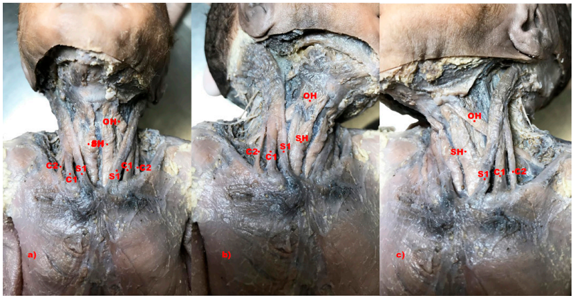
S, sternal head; C, clavicular head; SH, sternohyoid muscle; OH, omohyoid muscle; Numbers 1-2 indicate the heads; SCM, sternocleidomastoid muscle
The sternocleidomastoid muscle (SCM), located in the anterolateral region of the neck, serves as an important mark on the division of the anterior and posterior triangles. It originates from the sternum and the clavicule, and is inserted on the temporal bone mastoid process.1 The SCM is one of the most complex muscles of the body, it is responsible for the mechanical action of most head movements and is considered an accessory muscle for breathing.2 Usually the SCM has two heads, one sternal and the other clavicular. The sternal head emerges from the anterior face of the sternum manubrium as a distinct rounded tendon of consistent width, whereas the clavicular head covers the clavicule medial third and can vary in width, emerging as a band shaped tendon with musculofibrous appearance.3 The SCM is composed of five parts disposed in two layers: a superficial one, composed by the superficial part of the sternocleidomastoid, sternooccipital and cleidooccipital, and a deep one, that consists of the deep part of the sternomastoid and the cleidomastoid part.4 However, several variations can occur, including one or more accessory heads.5–7 Therefore, it is of great importance that the doctors have a clear comprehension of the SCM anatomy and its possible variations, in order to prevent inadvertent complications. The goal of this study was to report the presence of the bilateral three headed sternocleidomastoid muscle.
During dissection routine, on the Anatomy Laboratory of the Federal University of Sergipe, of a 23,9 weeks old male human fetus cadaver, after the removal of the superficial cervical fascia and the platysma muscle, were observed bilateral supernumerary heads on the sternocleidomastoid muscles (Figure 1A). On both sides the muscle had an additional head which originated in the medial third of the clavicular portion (Figure 1B, 1C). Both clavicular heads of the sternocleidomastoid muscle were separated by a wider triangular space, when compared to the triangle formed between the usual sternal and clavicular heads, which corresponds to one more superficial depression, the additional minor supraclavicular fossa. Both on the right side as on the left, the sternal heads originated from the superolateral parts of the sternum manubrium and the capsules of sternoclavicular joints, and they were separated from each other by a distance of 4,13mm.

Figure 1 Photograph showing, bilaterally, the supernumerary heads of the sternocleidomastoid muscle (a) on the right side (b) and on the left side (c) of the neck
Abbreviations: S, sternal head; C, clavicular head; SH, sternohyoid muscle; OH, omohyoid muscle; Numbers 1-2 indicate the heads
On the right side, the three heads united at the level of the hyoid bone, with a distance of approximately 22,65mm from the origin of the heads, its total length was of 46 mm and its width in the insertion was 14,06mm and extended itself from the temporal bone mastoid process to the lateral third of the upper nuchal line. And on the left side, this union of the heads occurred at 20,22mm from the heads origin, also at the level of the hyoid bone and its total length was of 40mm, with 17,13mm of insertion width that also extended from the mastoid process to the lateral third of the upper nuchal line. The width, on the right side, of the sternal head insertion was 3,41mm, the medial clavicular head was 4,01mm and the lateral head was 2,10mm. Whereas, on the left side, the width of the sternal head was of 3,62mm, the medial clavicular head was 4,76mm and the lateral head was 1,8mm. The distance between the bases that formed the superficial triangles of the minor supraclavicular fossa was of 1,27mm on the right side and 1,18mm on the additional minor supraclavicular fossa, on the left this distance was of 1,30mm and 0,60mm, respectively.
The frequency of the bilateral additional heads of the SCM is still more rare than unilateral variations of the head.8 However, the variations on its insertions are even rarer.9 In our report, an additional clavicular head was observed bilaterally on each side of the body, and it directed obliquely towards the temporal bone mastoid process and the upper nuchal line, which was also observed by Ramesh et al.10 and Anil et al.11 on a male cadaver. This supernumerary heads variations of the SCM can be caused by fusion failure or abnormal mesodermal division during the development.12
Boaro Fragoso13 reported the presence of three clavicular heads on a nine months old infant. Natsis et al.9 observed bilateral supernumerary heads of the SCM that had an additional sternal head and three additional clavicular heads, totalizing six heads (including the usual SCM heads). Kaur et al.14 also observed that the right SCM was formed by six heads, two sternal and four clavicular, while the left muscle did not present any variation. Besides these variations, similar to our case, several authors9,13–15 also noted that the minor supraclavicular triangle was very narrow, which is in accordance to our report, where the triangle which formed the additional minor supraclavicular fossa was also smaller. The knowledge of the anatomical variations of the SCM origin is very important to vascular and orthopedic surgeons, neurosurgeons, and especially anesthesiologists, who intervene on the minor supraclavicular fossa during the implantation of a central venous catheter, which could lead to pleural perforation and, consequently, pneumothorax.
Law n° 8.501, from the 30th of November of 1992. Provides for the use of unclaimed cadavers for the purposes of scientific studies or research and other aspects.
We would like to thank José Bispo da Silva and Luis Henrique Santos Fortes, technicians from the anatomy laboratory, for their support in preparing of the body. .
Author declares there are no conflicts of interest.
None.

©2020 Aragão, et al. This is an open access article distributed under the terms of the, which permits unrestricted use, distribution, and build upon your work non-commercially.