MOJ
eISSN: 2471-139X


Research Article Volume 11 Issue 1
1Department of Human Anatomy, School of Medicine, Maseno University, P.O. Box 333-40105, private bag Maseno, Kenya
2Department of Dentistry, School of Medicine, Maseno University, P.O. Box 333-40105, private bag Maseno, Kenya
Correspondence: Ode Brian Odhiambo, Department of Human Anatomy, School of Medicine, Maseno University, P.O. Box 333-40105, private bag Maseno, Kenya
Received: April 18, 2024 | Published: May 24, 2024
Citation: Odhiambo OB, Opondo I, Adero W, et al. Age estimation using orthopantomograms and Willems method among Kenyan children attending dental clinics in Western Kenya. MOJ Anat Physiol. 2024;11(1):15-18. DOI: 10.15406/mojap.2024.11.00343
Background: Various methods have been used to estimate age in different populations among them being Willems method which has widely been utilized. In Kenya, there is hardly any approved method that has been used to achieve this purpose, hence the need to determine the available methods for estimating age of children in Western Kenya. Therefore, this study aimed at estimating age using Willems method among children attending dental clinics in Western Kenya.
Methods: The study adopted a cross-sectional descriptive design and used Yamane Taro (1967) formulae to find a sample size of 171 orthopantomograms (94males and 77 females) out of 300 panoramic radiographs of children aged between 5-17 years. They were examined by the author in order to determine the tooth maturity stages (A-H) for the first seven mandibular teeth on the left side, accorded maturity scores according to Willems conversion tables for boys and girls and summed up to obtain dental age. Descriptive and inferential statistics were used to analyze data. The results were then presented in tables and figures.
Results: The overall mean dental age was 8.94 ± 2.64 with a standard error of 0.173 and that of females and males was 8.75 ±2.28 and 9.10 ±2.24 years respectively at 95% Confidence interval.
Conclusion: In conclusion, Willems method revealed an overall underestimation of dental age with no statistical difference between estimated and actual age in both genders among children in Western Kenya.
Keywords: chronological age, dental age, orthopantomograms, Willems method
Age forms an important part of an individual’s biodata that is not only necessary for the living but also for the deceased. Birth registration documents such as certificate of birth, national identity cards or passports can be used to verify an individual’s age. Nevertheless, in some circumstances where an individual’s age can’t be established, age estimation must be authenticated.1 Age estimation is normally applied in diverse fields such as forensic medicine, odontology, anthropology and archeological studies.2–4 In this regard, it is important to understand that Chronological age (CA), also known as actual age, is obtained from the birth certificate of a newborn child while Dental age (DA) or estimated age is obtained by looking at the growth and development of individual’ teeth.5 Several methods have been used to estimate age in different populations. Among the most widely used is Willems method which was based on Belgian Caucasian reference population. Willems applied part of Demirjian (1973) method by using the same A-H tooth staging technique relying on the left seven mandibular teeth. Once the stages of development for the seven permanent teeth have been identified, each tooth stage is then accorded new maturity scores and summed up to obtain dental age using Willems conversion tables for boys and girls. In Belgian Caucasian population,6 Willems used Demirjian’s method to estimate age. The study gave out a mean chronological and dental age difference of 0.5 & 0.9 years for male and female children respectively showing an overestimation. As opposed to other methods, Willems method was confirmed to be more accurate with the smallest mean and standard deviation in age estimation. In Macedonian children, Willem’s method evidently performed better than Demirjian.7 Among the Egyptian population, it performed better as opposed to Cameriere method with a difference of -0.15 years & -0.29 years respectively in terms of mean.8 In Kenya therefore, the study seeks to test the performance of Willems method in age estimation of children attending dental clinics in Western Kenya due to few established national standards for age estimation, few publications on dental maturity and utilization of scoring radiographic age estimation methods in this region.
This was a cross-sectional retrospective study conducted in a dental clinic and approved by different authorities (MSC/SM/ 00011/020, MUSERC/01149/22, and NACOSTI/P/22/2240) and concerned ethical committees. The study targeted digital orthopantomograms of Kenyan children aged between five to seventeen (5-17 years) from dental clinic records. This age bracket is a recommendation of the American Dental Association Council on Scientific Affairs (2006) where Panoramic radiographs are considered for children with an evidence of permanent tooth eruption which likely occurs at 5-7 years. The maximum age limit for this study was 17 years as this is the average age where adolescents attain full teeth maturity as evidenced by the third molar growth. This study adopted a cross-sectional descriptive design and used Yamane Taro (1967) to obtain a sample of 171 panoramic radiographs (94 males and 77 females) from a total of 300 radiographs. Purposive sampling method was used to select the sample radiographs. Data collection form was pre-tested and checked for completeness to minimize errors. Any omitted data was rechecked and entered. The radiographs in soft copy were retrieved from a computer data base connected to the digital panoramic x-ray machine by the research assistants from the dental clinic. The digital orthopantomograms was a product of GENDEX ORTHORALIX 9200 machine with all the standard protocols in place. The date of panoramic imaging (DOP) and date of birth (DOB) was noted for each radiograph and every image coded and assigned Arabic numerals (1 M, 2M for males…and 1F, 2F for females) in order to conceal any identity of the children and avoid biasness. Chronological age was calculated by subtracting the Date of birth (DOB) from Date of panoramic imaging (DOP) i.e. (DOB-DOP) and expressed in two decimal points. The ages of children were then grouped into six age cohorts i.e. From (5-6.99 years) up to (15-17.99 years). The inclusion criteria included; radiographs with quality diagnostic images, no missing teeth on the mandibular segment, and those with available information on the date of birth and date of panoramic imaging. The radiographs that had missing biodata, and those with pathologies and cysts on teeth dentition were excluded. The panoramic radiographs were then examined by looking at the morphological appearance of the teeth and staged (A-H) according to Demirjian tooth staging technique. The technique was applied on the seven mandibular left teeth and each maturity stage was accorded maturity scores, summed up, and converted to dental age according to the Willems conversion tables for boys and girls. The data was then put into an Excel sheet and uploaded into the statistical package for social sciences (SPSS) version 26.0. Descriptive statistics such as mean, standard deviation, minimum and maximum was utilized and presented in tables and figures while inferential statistics such as standard error of mean and linear regression were used to measure the deviation and statistical significance. 10% of the images were selected randomly to measure intraexaminer reliability. Content Validity was verified by an expert from the paediatric dental department.
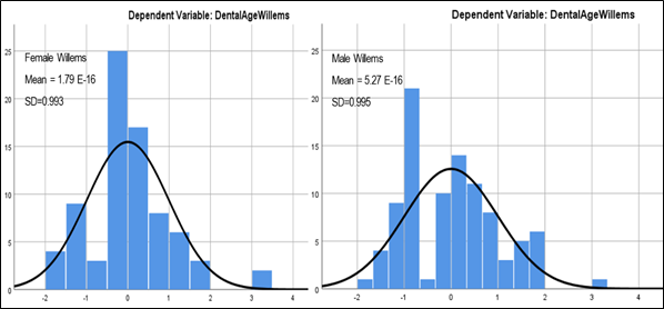
Using Willems method, the mean dental age was 8.94 ± 2.264 with a standard error of the mean of 0.173 in the total respondents (Table 1.1). In females, the mean dental age was 8.75± 2.289 with a standard error of mean of 0.261 while in males it was 9.10±2.244 with a standard error of mean of 0.231(Table 1.2). The probability of underestimation of chronological age was therefore high in females than in males. However, the cumulative error for age estimation for both sexes was less than ± 1.95 years (Table 1.3). Using the regression analysis test for linearity, the mean difference between chronological age and estimated age was plotted against the age frequency distribution to determine how wide the deviation is from the chronological age (Figure 1.1). Among females, the deviation from chronological age was ±2.062 years at 95% confidence interval (CI), while in males, the deviation was ±1.952. This means that the females had a wider margin of error during age estimation than males (Table 1.3) (Figure 1).
Estimated dental age using Willem’s method
Using Willems method, the mean estimated age of all the respondents was 8.94± 2.264 with a standard error of 0.173 years (Table 1.1).
In females, the mean estimated age using Willems method was 8.75± 2.289 with a standard error of mean of 0.261. Among the males, the mean estimated age was 9.10±2.244 with a standard error of 0.231 (Table 1.2).
The distribution frequency of dental age estimation errors is illustrated in Figure 1.1 using Willems method. The chronological age of 57% of females were underestimated while 56% of the males were underestimated using Willems method of age estimation. Among females, the deviation from CA was ±2.062 years at 95% confidence interval (CI), while in males, the deviation from CA was ±1.952. The probability of underestimation of CA was therefore high in females using Willems method. However, the cumulative error for age estimation for both sexes was less than ± 1.95 years (Figure 1.1 and Table 1.3).
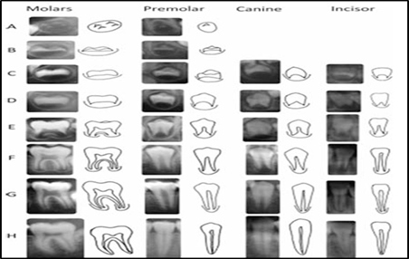
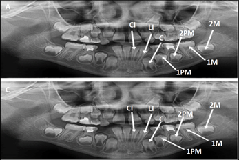
Willems method is a modified Demirjian (1973) where scoring was done using same A-H staging technique relying on the left seven mandibular teeth. Once the stages of development for the seven permanent teeth were identified then each tooth stage was accorded new maturity scores according to the Willems conversion tables and summed up to obtain dental age6 (Figure 1.2 & 1.3). Studies done in various populations have found Willems method to either Under or overestimate the dental age (DA) with an average of 0.6 years (7.2 months or less).9,10 In the present study, there was an overall underestimation of mean dental age at 8.94 ±2.264 with a standard error of 0.173 years (Table 1.1). Similar observations made among 946 children aged 3-16.99 years from Bangladesh and British Caucasian ethnic origin revealed an overall underestimation of 8.02±0.93.11
The mean dental age was 8.75±2.289 in females and 9.10±2. 244 in males using Willems method (Table 1.2). In the entire age cohort, the difference in estimated dental age varied from 0.000-1.557 in females and 0.447-1.373 in males (Table 1.2). The greatest underestimation in females was found among the cohort aged 11-12.99 years followed by 15-17.99 years and 5-6.99-years. In the males, the greatest underestimation was found in the 9-10.99 followed by 7-8.99- and 5-6.99-year-old age groups (Table 1.1). In addition, before the age of 10 years, the males were more advanced in their dental age compared to the females who took the lead after 10 years (Table 1.2)These findings were also similar to those observed in south Indian children aged 3-15 years where the greatest underestimation in females was found in 8-9.99 years followed by 14-15.99 years and 12-13.99-year-old age groups while in males, the greatest underestimation was found in 14-15.99 years followed by 12-13.99- and 8-9.99-year-old age groups. In the contrary, this population (South Indian children) had an overestimation in the 10-11.99-year-old age group.
In the present study, it was noted that Willems showed a high probability of underestimation in females at 57% than in males at 56 % which is in agreement with studies conducted in North Indian children aged 6-15 years where Willems method underestimated chronological age of 58% females and 56% males.12 This is in contrast to studies done among 330 Turkish children aged 5-15 years where Willems performed better in males than females.13 Among females, the deviation from the chronological age was ±2.062 at 95% confidence while in males the deviation was ±1.95 (Figure 1.1 & Table 1.3). This depicted that females had a wider margin of error as opposed to males. Moreover, it was observed that there was a significant delay in the dental maturation of females and males similar to observations seen in South Indian children aged 6-14 years who had a delayed dental maturation of 0.08 years and 0.69 years in females and males respectively. The delay in dental maturity maybe partly explained by population differences, genetic variations, nutritional factors, socio-economic status, dietary habits and lifestyle. Therefore, Willems method was found to perform better in females than in males in age estimation.
|
Dental Age, Willems |
|||||||
|
Age cohort |
N |
Mean |
Std. Deviation |
Std. Error of Mean |
Minimum |
Maximum |
Variance |
|
5-6.99 |
25 |
6.12 |
1.13 |
0.226 |
5 |
9 |
1.277 |
|
7-8.99 |
47 |
7.62 |
1.171 |
0.171 |
7 |
11 |
1.372 |
|
9-10.99 |
49 |
9.08 |
1.152 |
0.165 |
9 |
14 |
1.327 |
|
11-12.99 |
29 |
10.52 |
1.153 |
0.214 |
10 |
15 |
1.33 |
|
13-14.99 |
12 |
12.33 |
0.492 |
0.142 |
13 |
14 |
0.242 |
|
15-17.99 |
9 |
13.33 |
1 |
0.333 |
13 |
16 |
1 |
|
Total |
171 |
8.94 |
2.264 |
0.173 |
5 |
16 |
5.126 |
Table 1.1 Dental age using Willems method in the total sample population
|
Female Willem’s |
Male Willems |
|||||||
|
Age cohort |
N |
Mean |
SD |
SEM |
N |
Mean |
SD |
SEM |
|
5- 6.99 |
13 |
5.77 |
1.092 |
0.303 |
12 |
6.5 |
1.087 |
0.314 |
|
7- 8.99 |
21 |
7.86 |
0.91 |
0.199 |
26 |
7.42 |
1.332 |
0.261 |
|
9 -10.99 |
23 |
8.87 |
0.815 |
0.17 |
26 |
9.27 |
1.373 |
0.269 |
|
11-12.99 |
13 |
10.62 |
1.557 |
0.432 |
16 |
10.44 |
0.727 |
0.182 |
|
13 -14.99 |
3 |
12 |
0 |
0 |
9 |
12.44 |
0.527 |
0.176 |
|
15 -17.99 |
4 |
14 |
1.155 |
0.577 |
5 |
12.8 |
0.447 |
0.2 |
|
Total |
77 |
8.75 |
2.289 |
0.261 |
94 |
9.1 |
2.244 |
0.231 |
Table 1.2 Estimated dental age using Willem’s method in males and females
|
Females |
Males |
|||||
|
Predicted models |
Minimum |
Maximum |
Std. Deviation |
Minimum |
Maximum |
Std. Deviation |
|
Predicted Value |
5.6 |
14.76 |
2.062 |
5.93 |
14.48 |
1.952 |
|
Residual |
-1.944 |
3.056 |
0.994 |
-2.064 |
3.51 |
1.107 |
|
Std. Predicted Value |
-1.529 |
2.913 |
1 |
-1.624 |
2.759 |
1 |
|
Std. Residual |
-1.944 |
3.055 |
0.993 |
-1.855 |
3.155 |
0.995 |
Table 1.3 Linear regression test for relationship between chronological age and Dental age in males and females

Figure 1.1 Histogram illustrating error in Dental age estimation in boys and girls using Willems method.

Figure 1.2 Demirjian stages of tooth development(A-H).
Adopted from the assessment and interpretation of Demirjian dental maturity stages by Hellen M. Liversidge, (2010).

Figure 1.3 Digital orthopantomogram images of 12 year -old male and female child whose Dental ages were estimated at 10 and 9 years respectively using Willems method. (A & B).
Key: CI-Central incisor, LI -Lateral incisors, C- Canines, 1PM- First Premolar, 2PM- Second Premolar, 1M –First Molar, 2PM- Second Molar.
The current study findings suggested an overall underestimation of the dental age in both females and males however, Willems method performed better in females which had the highest probability of age underestimation and a wider margin of error.
I acknowledge my co-authors, Dr. Immaculate Opondo, Dr. Walter Adero, Dr. Willis Oyieko and Dr. Domnic Marera for their immense support and advice towards my manuscript writing. My appreciation also extends to Maseno University and specifically School of Medicine and Human Anatomy Department for offering me an opportunity to pursue my Master’s degree in Human Anatomy. Lastly, I would like to acknowledge other lecturers from the postgraduate school and my colleagues who played an important role in the journey of my postgraduate studies.
No consent was needed.
No conflict of interest.

©2024 Odhiambo, et al. This is an open access article distributed under the terms of the, which permits unrestricted use, distribution, and build upon your work non-commercially.