MOJ
eISSN: 2471-139X


Mini Review Volume 4 Issue 4
1Department of Anatomy and Cellular Biology, University of Baghdad, Iraq
2Novel Psychoactive Substances research unit, University of Hertfordshire, UK
Correspondence: Ahmed Al Imam, House 18/5, Al-Akhtal Street, District 318, Al-Adhamyia, 10053, Baghdad, Iraq
Received: September 03, 2017 | Published: November 1, 2017
Citation: Al-Imam A, Al-Hadithi N. A biomedical device innovation: a method for accurate calculation of volume (volumetry) of irregular bone specimens. MOJ Anat Physiol. 2017;4(4):327–329. DOI: 10.15406/mojap.2017.04.00141
The morphometric studies are considered to be abundant in connection with the study of Human Anatomy and Comparative Anatomy. The estimation of volume, also known as volumetry, is integral to these studies although it is not frequently included. Several methods exist to measure the accurate size (volume) of bones and certain soft tissue; these techniques are not limited to replica manufacturing and the implementation of Archimedes’ principles of buoyancy and fluid displacement. The latter will be explored in connection with a prospective biomedical innovation of a device for the purpose of estimation of the size of bones particularly those of variable shapes, dimensions, and highly irregular shapes.
Keywords: biomedical engineering, equipment and supplies, osteology, morphometry, volumetry, hydraulics, casting, cast materials, replica techniques, innovation, invention
There has been an abundance of research efforts that are pertinent to the disciplines of Human Anatomy, Veterinary Anatomy, Anatomy of vertebrates, and Comparative Anatomy; these were based on gross (macrosopic) examination of tissues including bones and soft tissue. Numerous studies were based on morphometric analyses of relevance to bones specimens (osteology-based).1‒5 The morphometric studies of bones were usually focusing on observing and quantifying; morphologies and shapes, dimensional parameters, surface area of articular and non-articular regions, and volumetric measurements (volumetry) of a whole bone or segments of bone (processes, tubercles, tuberosities, cavities, sinuses, and others).2,5‒8
The measurement of the volume of a bone or a segment of bone, also known as volumetry, is an integral component of morphometric studies; it can also be applied to cartilaginous tissues and other chemically-treated (processed) soft tissues of interest. Not all morphometric studies employ the use of volumetric analysis despite its vital importance; in addition, several morphometric studies also neglected the analysis and quantification of surface areas.6‒8 All these aspects can lead to weak studies in connection with morphometry and can result in low level-of-evidence when assessed by critical appraisal tools and instruments of Evidence-based Medicine.9
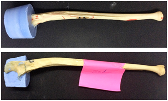
There are some methods that are frequently utilized for the purpose of volumetry; the most common techniques include the use of cast impression materials and replica techniques (Figure 1) (Figure 2), the utilization of liquids with particular surface tension and specific hydrophobic-philic properties to measure the volume of cavities and impressions, and the exploitation of Archimedes' principles of buoyancy and fluid displacement.5‒8,10‒12 In this article, we explore the concept of a biomedical innovation, a concept of a device, that is being designed for the purpose of accurate measurements of volumes; the device is to be made of a transparent cylindrical container of a pre-specified diameter, length, and volume; the measurement of volume of bones and soft tissue should rely on the Archimedes' principles of buoyancy and fluid displacement. Further, the cylinder can be calibrated in accordance with the type and size of the specimen to be studied. On the other hand, this device should give an accurate electronic quantification of the size of bone samples immersed in it; the device can be based on some physical principles including; light amplification by stimulated emission of radiation (LASER), galvanic and electronic conduction, or an electronic vernier caliber in combination with basic mathematic formulae for the calculation of volume.7,13‒15

Figure 1 Cast material (alginate impression material) for calculation of volume of the proximal segment of ulna.
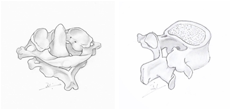
The concept of this device, as simple as it is, can revolutionize all of the volumetry-based morphometric studies; it can be used for bones of variable shapes and dimensions including those of minute size as in the case of the middle ear ossicles, and can be applied even to bones of irregular shapes including those of the carpal bones, tarsal bones, ribs, and vertebrae (Figure 3). Further, it can be used for soft tissues including the intervertebral discs, symphysial tissues, and interarticular discs; it can also be utilized to measure inner bone cavities (medullary and marrow cavities), surface projections and impressions as in the case of the mandibular (glenoid) fossa, and even to cavities within bones, for instance, the carotid canal within the petrous part of the temporal bone and the sphenoidal air sinus within the body of the sphenoid bone of the cranium. Other soft tissues can also be measured, after being chemically processed to fixed (mummified) volumes, including those of joints, glands, and small muscles of the head and neck, for example, the infrahyoid muscles, pterygoid muscles, muscles of the styloid apparatus, muscles of the inner ear, and the extraocular muscles. Hence, the (bio) medical applications can be endless in connection with the study of Anatomical Sciences. Ω Concept Art by Shams Al-Nuaimi.

Figure 3 Posterolateral view of some irregular bones: Atlanto-axial Articulation (left) and Typical Lumbar Vertebra (right).
This technique should also be favourable and far more accurate than other methods implemented for volumetry, including casting and replica manufacturing; our innovative approach will be more precise, less time-consuming, and requires simple efforts, and can be carried out with little training unlike model (replica) manufacturing which usually necessitates a reasonable training and expertise to reach out accurate results (estimation of volumes). However, there are some restrictions on this prospective device; eroded bones can give rise to an erroneous estimate of size (volume). Similarly, the presence of nutrient foramina as these will take up some liquid while being immersed in it; these two obstacles can be overcome via the use of specific hydrophobic paint materials to be applied to the surfaces of these bones. Other enhancements may include the use of liquids (for immersion of bones) of low surface tension, calibration(s) peculiar to the type and size-shape of the bone to be measured, the use of chromatic liquids, and double checking of the results with other techniques of volumetry including replica manufacturing.
The currently utilized methods for calculation of the volume of osteological and non-osteological specimens are either primitive, crude, time and efforts-consuming or lacks high accuracy. We propose an innovative approach for a precise electronic estimation of the volume of tissues of interest especially those of bones of highly irregular shapes. Further, this engineered technique can be implemented in combination with other methods, including replica manufacturing, for an accurate volumetric measurement of cavities within the bone, in between bones and tissues, and surface impressions or projections belonging to these tissues. The aim is to provide researchers with easier means for volumetric assessment while conserving the resources, both financial and manpower, including the prerequisite requirements for advanced training and expertise for making casts and replica manufacturing techniques.
The authors would like to acknowledge the efforts of Shams Al-Nuaimi for the provision of the illustration of cervical and lumbar vertebrae (Figure 3); Shams is a third-year medical student at the College of Medicine-University of Baghdad in Iraq.
The author has nothing to be declared.
This study was entirely self-funded.

©2017 Al-Imam, et al. This is an open access article distributed under the terms of the, which permits unrestricted use, distribution, and build upon your work non-commercially.