Journal of
eISSN: 2475-5540


Case Report Volume 2 Issue 5
Department of Pathology, University of Iowa, USA
Correspondence: Chen Zhao, Department of Pathology, University of Iowa, 25 S. Grand Avenue, 1163 ML, Iowa City, Iowa 52242-1181, USA, Tel 3193844668, Fax 3193358453
Received: April 21, 2017 | Published: April 26, 2017
Citation: Schouweiler K, Zhao C. CD5+ diffuse large b-cell lymphoma mimicking metastatic carcinoma to bone marrow. J Stem Cell Res Ther. 2017;2(5):136. DOI: 10.15406/jsrt.2017.02.00074
carcinoma, b-cell lymphoma, cd5, lymphadenopathy
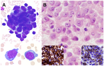
An 89-year old man with a history of prostatic adenocarcinoma presents with fatigue, mild splenomegaly and lymphadenopathy, and pancytopenia. The bone marrow biopsy was received for consultation from an outside institution. The aspirate smears contain scant, largely acellular particles and scattered grape-like clusters of large atypical cells (A, upper panel). The atypical cells have irregular centrally to eccentrically placed nuclei, relatively fine chromatin, distinct nucleoli and variable amounts of pale blue cytoplasm (A, lower panel). Flow cytometric analysis of the marrow aspirate shows no increased blasts and no atypical myeloid or lymphoid populations.
Sections of the core biopsy and clot show a hypocellular bone marrow (<10%) with decreased trilineage hematopoiesis. Multifocal infiltrative clusters of atypical pleomorphic cells are present and show morphology similar to that observed on the aspirate (B). Immunohistochemical staining of the atypical cells on the core biopsy is positive for CD45, CD79a and CD20 (B, left inset); a major subset is positive for CD5 (B, right inset) and a minor subset is positive for MUM-1. Staining with PAX5, CD10, CD56, CD34, ALK-1, CD138, CD30, CD3, BCL-6, TdT, cyclin D1, MPO, and CD163 is negative. Kappa and Lambda light chain staining is non-contributory. Additional immunohistochemical staining for AE1/3, CAM5.2, PSA, PSAP and S100 is negative (Figure 1).

In this case, flow cytometry was unremarkable and the clinical history and aspirate morphology (cohesive cell clusters) were highly suggestive of involvement by metastatic carcinoma. The final diagnosis was determined to be CD5+ diffuse large B-cell lymphoma (DLBCL) with bone marrow involvement. It should be noted that CD5+DLBCL has a poor prognosis, which requires intensive treatment.1,2 The unusual cohesive growth pattern of DLBCL is only rarely reported in the literature.3,4 While morphology is an important aspect of diagnosis, failure to definitively exclude hematopoietic mimics of carcinoma, particularly in the setting of epithelioid morphology with unremarkable flow cytometry, may hinder a correct diagnosis.
None.
The author declares no conflict of interest.

©2017 Schouweiler, et al. This is an open access article distributed under the terms of the, which permits unrestricted use, distribution, and build upon your work non-commercially.