Journal of
eISSN: 2373-4426


Case Report Volume 14 Issue 3
1Consultant Radiologist, Department of Radiodiagnosis, MGS Super speciality Hospital, India
2Post Graduate Resident (NBEMS), Guru Gobind Singh Govt Hospital, Government of Delhi, India
3Consultant Pediatrician & Neonatologist, Department of Pediatrics, Guru Gobind Singh Government Hospital, Government of Delhi, India
Correspondence: Dr. Abhinav Gupta, Department of Radiodiagnosis, MGS Super speciality Hospital, Punjabi Bagh, New Delhi, Delhi-110026, India, Tel +919891070040
Received: November 20, 2024 | Published: December 19, 2024
Citation: Gupta A, Vemula S, Gupta AK, et al. Traumatic separation of distal femoral epiphysis in a neonate following breech delivery: a case report. J Pediatr Neonatal Care. 2024;14(3):212-213. DOI: 10.15406/jpnc.2024.14.00569
This case report describes a male neonate delivered by an emergency caesarean section at 34 weeks of gestation, weighing 1800 grams. The mother was an un-booked case and presented directly in labor with a footling breech. The extraction of the baby was difficult. Despite this, the neonate had a normal Apgar score and was transferred to the special care neonatal unit with mild respiratory distress. Since maternal history included prolonged rupture of membranes, the neonate was initiated on intravenous prophylactic antibiotics. On the 3rd day of life, the neonate developed swelling and restricted movement of the left knee. Initial evaluation, including orthopedic consultation, raised suspicion of septic arthritis, but synovial fluid analysis was negative. Plain radiography on day 5 of life revealed traumatic separation of the distal femoral epiphysis. The fracture was reduced by an orthopedic surgeon by closed reduction, and a cast was applied for 4 weeks. The baby was referred to a higher center for further management. This case highlights the importance of considering this rare complication in neonates following breech delivery.
Traumatic separation of the distal femoral epiphysis, also termed distal femoral epiphysiolysis (DFE) is a rare complication of breech delivery in neonates, especially challenging to diagnose without ossification of the distal femoral epiphysis, particularly in preterm neonates. However, in this pre-term neonate, radiographic findings were diagnostic, emphasizing the importance of considering this diagnosis in neonates with knee swelling after breech delivery.
A male neonate was delivered by emergency cesarean section at 34 weeks of gestation, weighing 1800 grams. The extraction was challenging due to the mother's late presentation in labor with a breech presentation, specifically a footling presentation. Initially, attempts were made to extract the baby by pulling the leg, but this was unsuccessful, finally, the baby was extracted from the buttocks. Despite the difficult extraction, the neonate cried promptly after birth and had a normal Apgar score of 7 and 8 at 1 and 5 minutes, respectively. The baby was transferred to the special care neonatal unit with a diagnosis of prematurity with mild respiratory distress. With a history of prolonged rupture of membranes for more than 48 hours, the baby was initiated on intravenous ampicillin and gentamycin as per hospital protocol. A complete blood count (CBC) at 6 hours of age showed normal results, and C-reactive protein (CRP) was negative. The respiratory distress resolved within 24 hours.
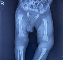
On the 3rd day of life, the neonate developed swelling of the left knee, including the lower thigh, along with restricted movement of the left leg, with the knee kept flexed and mildly tender. There were no signs of local inflammation or skin changes over the affected knee, and the local temperature over the swelling was not raised. A repeat CBC at 48 hours of age showed normal leukocyte counts, and CRP was non-reactive. Plain radiography of both knee joints revealed a wide joint space on the left side with the absent distal femoral epiphysis, contrary to the presence of the right femoral distal epiphysis visible due to the ossification of its center. However, despite the non-visualization of the distal femoral epiphysis on the left side, the femur and tibia/fibula on the affected side appeared to align normally (Figure 1); hence birth injury could not be suspected initially. The orthopedic surgeon suspected septic arthritis and aspirated a small amount of synovial fluid from the left knee joint. On evaluation, the fluid was clear, negative for pus cells, and the gram stain. The bacterial culture of synovial fluid was also negative after 24 hours of incubation.

Figure 1 Anteroposterior radiograph of both knee joints on day 3 of life showing a wide joint space on the left side with the absent distal femoral epiphysis.
A mild increase in left knee swelling was noticed on day 4; eventually, antibiotics were upgraded to intravenous cefotaxime and amikacin empirically. However, the baby was afebrile and clinically not sick but was in pain on passive movements of the left lower limb.
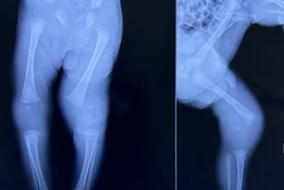
Considering the negative tests for sepsis and septic arthritis and persistence of, and slight increase in, knee swelling, plain radiographs of both knee joints were repeated on day 5 of life. The radiographs revealed swelling and a wide joint space in the left knee joint, along with valgus deformity of the femur. Surprisingly, the distal femoral epiphysis, visible by its ossified center, was seen to have lost alignment with the femur shaft but continued its alignment with the tibia (Figure 2), confirming the diagnosis of distal femoral epiphysiolysis.
The fracture was reduced by an orthopedic surgeon by closed reduction, and a cast was applied for 4 weeks. The baby was referred to higher center for further management, and follow-up in view of the risk of long-term shortening of the affected limb.

Figure 2 Anteroposterior radiograph of both knee joints and lateral radiograph of left knee joint on day -5 of life showing a wide joint space in the left knee joint, along with valgus deformity of the femur. The distal femoral epiphysis, visible by its ossified center, has lost alignment with the femur shaft but continued its alignment with the tibia.
The distal femoral epiphysis (DFE) is the first of the long bone epiphyses to ossify and appear before the proximal tibial epiphysis. Around birth, the DFE consists of a rounded or oval nucleus with pitting and osseous circumvolutions.1
Traumatic separation of distal femoral epiphysis or DFE in newborn infants is rare.2–7 This can result from difficult extraction of lower limbs in breech delivery.2–4 However, some cases have been reported even after the extraction of baby with breech presentation during caesarean section,5 like in the present case.
Diagnosis of a DFE in the newborn may be difficult as the condition may be missed on conventional radiographs. Moreover, with clinical signs of inflammation the condition may remain undiagnosed or misdiagnosed as septic arthritis or physeal osteomyelitis. Delayed diagnosis of epiphyseal fractures of the distal femur may lead malunion and deformity. If untreated may lead to growth arrest.2–6
Advanced imaging techniques like ultrasonography, CT, and MRI can help improve diagnostic accuracy.6,7 Fortunately, the distal femoral epiphysis in the present case despite being a preterm neonate, showed an ossified center visible on x-ray, enabling its diagnosis through conventional radiographs.
Traumatic separation of the distal femoral epiphysis is a rare complication of breech delivery and challenging to diagnose. This case underscores the importance of considering this diagnosis in neonates with knee swelling following breech delivery.
None.
This research received no external funding.
The authors declare no conflicts of interest.

©2024 Gupta, et al. This is an open access article distributed under the terms of the, which permits unrestricted use, distribution, and build upon your work non-commercially.