Journal of
eISSN: 2373-4426


Mini Review Volume 6 Issue 1
1Department of Surgery, All India Institute of Medical sciences Raipur, India
2Department of Pediatric Surgery, King George?s Medical University, India
3Department of Pediatric Surgery, All India Institute of Medical sciences, India
Correspondence: Sunita Singh (MS, MCh, paediatric surgery), Assistant Prof. Dept of Surgery, All India Institute of Medical Sciences, Raipur, Chhattisgarh, India, Tel 8518887725
Received: October 31, 2016 | Published: January 3, 2017
Citation: DOI: 10.15406/jpnc.2017.06.00229
Twelve % of Wilms’ tumor patients have metastases at initial presentation. The hematogenous metastasis is most common in lung, followed by liver, contralateral kidney, bone and brain. In the literature (1928-2016) 12 cases of testicular/ paratesticular metastasis of Wilms’ tumor has been reported. Here, we report a child having Wilms’ tumor with testicular metastasis, with discussion on how to approach the children having simultaneous renal and testicular mass.
Keywords: metastastic wilms’ tumor; patent processes vaginalis; pediatric renal lump; Testicular metastasis; wilms’ tumor
AFP, alfa feto protein, CECT, contrast enhanced computer tomography, chemo: Chemotherapy; hCG, human chorionic gonadotropin hormone, HIO, high inguinal orchiectomy, i.e, that is; LDH 1, lactate dehydrogenase isoenzyme 1; RT, radiotherapy; SIOP, societe internationale d'oncologie pediatrique, WT, wilms’ tumor
Wilms’ tumor (WT)/ Nephroblastoma is the second common abdominal malignancy and most common malignant renal tumor in children.1 Twelve % of WT patients have metastases at initial presentation.2 Common hematogenous metastatic site of WT include the lung, liver, and contralateral kidney. Less common sites include the bone, skin, brain, and orbit.2 The lung is most common hematogenous metastatic site. The lymphatic spreads to lymph nodes are the most common. The rare site of metastasis are mediastinum and testis.1–12
In adults most frequent primary tumors metastatic to spermatic cord and epididymis are carcinomas from the stomach, prostate, colon, pancreas, appendix, and renal (renal cell carcinoma.13,14
It is utmost important to evaluate the genitalia in pediatric patients presenting with renal lump, as there may be syndromic association, (cryptorchidism, pseudohermaphroditism), vericocele (tumor compressing testicular veins) hydrocele (subclinical metastasis having reactive hydrocele ), patent processes vaginalis etc.15,16 To the best of our search (all languages, both indexed non indexed journal) with the key words testicular metastasis, paratesticular metastasis, metastatic Wilms’ tumor in the literature from 1928 to 2016, we found 12 cases of testicular/ paratesticular metastasis of WT.1–12 We excluded the cases of extrarenal scrotal WT (scrotal WT without primary renal involvement) arising from heterotropc anlage.17 Author here reported a rare case of WT with synchronous epsilateral testicular metastasis, with discussion of diagnostic and therapeutic approach for children presenting with renal lump and testicular mass.
A 3.5-year-old male was referred to us with left renal lump, gross haematuria, and a left testicular mass for 2 months. There was no history of fever, weight loss, cough, and hemoptesis etc. On examination a 12×8 cm, non-tender, hard, smooth, bimannualy palpable lump crossing the midline was present in left flank. Another 6.0×4.0 cm, well defined, smooth, non tender, hard, transilluminant negative left testicular mass was also present.
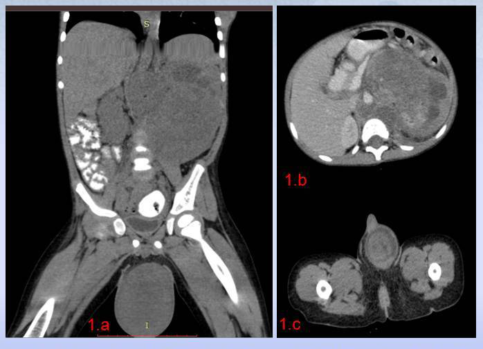
Blood biochemistory was normal except anemia. The serum Alfa Feto Protein (AFP), Humon Chorionic Gonadotropin (HCG) and Serum isoenzyme lactate dehydrogenase (LDH 1) were within normal limits. Ultrasonography suggested normal contralateral kidney and testicle. Contrast enhanced computer tomography (CECT) scan of abdomen with scrotum (shielding of right testicle) showed 16×10 cm left renal mass compressing adjacent structures, retroperitoneal lymph node enlargement, invasion of left renal vessels and inferior vena cava (below diaphragm) with a heterogenous 6×4.5 cm left testicular mass (Figure 1). Further radiology didn’t reveled any other site of metastasis.

Figure 1 1.a, b: 64 slice contrast enhanced CT scan of abdomen in coronal and axial section showing 16 ×10 cm sized heterogenous, left Renal mass, compressing adjacent structures, displacing inferior vena cava with multiple retroperitoneal lymph node enlargement and homogenous 6 ×4.5 cm sized left testicular mass; .1.c axial section of testicular mass showing homogenous testicular mass.
We adopted Societe Internationale D'oncologie Pediatrique (SIOP) protocol and through left transverse incision biopsy of left renal mass with hilar & para-aortic lymph node sampling was done.18 The procedure was accompanied by early control of left testicular vessels and radical orchiectomy through same incision. Histopathology showed blastemal type WT having diffuse anaplasia. The histology of left testis was same, with spermatic vessels involvement, and spared epididymis and spermatic cord. Hence we came to the diagnosis of testicular metastasis of primary WT.
We planned to administer the neoadjuvant chemotherapy according to SIOP stage III with high risk histology [19]. Vincristine, Adriamycin, Doxorubicin, Cyclophosphamide was administered with abdominal radiation therapy (RT). Five weeks after chemo-radio therapy gross haematurea settled down, but imaging didn’t showed regression in the size of renal or testicular mass. The chemotherapy shifted to etoposide, carboplatin and cyclophosphamide for 5 week with RT. The child was planned for surgery, but he lost the follow up.
The incidence of WT is 7.6 cases for every million children less than 15 years of age or 1 case per 10,000 infants.3 WT is associated with congenital syndromes in 10% of cases, including sporadic aniridia, isolated hemihypertrophy, Denys-Drash syndrome (nephropathy, renal failure, male pseudohermaphroditism, and Wilms’ tumor), genital anomalies, Beckwith-Wiedemann syndrome [visceromegaly, macroglossia, omphalocele, and hyperinsulinemic hypoglycemia in infancy and WAGR complex (WT with aniridia, genitourinary malformations, and mental retardation).15,16 This suggested a genetic predisposition to this tumor but literature analysis didn’t (Table 1) suggested any genetic/ familial predisposition for testicular metastasis.
|
S.No. |
Author’s |
Age |
Wilms’ tumor Side |
Testicular metastasis Side |
Presentation of testicular mass |
Treatment of testicular metastasis |
Histopathology of testicular SPECIMEN |
Outcome |
|
1 |
Dew et al.1 |
27 |
Lt |
Lt |
Testicular mass 6 m Post Radical Nephrectomy |
· HIO |
Testis, Epidydymis, spermatic cord, spermatic vein |
Died with pulmonary metastasis within 4 m of diagnosis testicular metastasis |
|
2 |
Yadav K et al.2 |
9 m |
Lt |
Lt |
Simultaneous renal and testicular mass |
· Biopsy Left renal mass and left radical orchiectomy through laparotomy incision · Chemo · Abdominal RT |
Spermatic cord |
Not available |
|
3 |
De Camereago et al.3 |
60 |
Rt |
Lt |
7 m Post Radical Nephrectomy (Hydrocele was present at index admission ) |
· HIO · Chemo · lung RT |
Epidydimis |
Lost Follow up |
|
4 |
Sauter et al.4 |
NA |
Lt |
Lt |
3 m Post Radical Nephrectomy |
· HIO · Chemo |
Testis, paratesticular soft tissue, spermatic cord, spermatic vein |
Not available |
|
5 |
Quattara et al.5 |
NA |
Rt |
Rt |
Massive bilateral testicular mass with small renal mass |
· Radical nephrectomy with bilateral partial orchectomy · Chemo · RT tumor bed and bilateral testis. |
Testis, spermatic vein |
Not available |
|
6 |
Trob et al.6 |
36 |
Rt |
Rt |
11 m Post Radical Nephrectomy (Hydrocele was present at index presentation) |
· HIO · Chemo |
testis |
NED 8.5 yr |
|
7 |
Aydin et al.7 |
36 |
Lt |
Rt paratesticular |
6 m Post Radical Nephrectomy (Hydrocele was present at index presentation) |
· HIO · Chemo · Abdominal RT |
Tunica vaginalis, tunica albuginea |
NED alive at 22 m |
|
8 |
Daher et al.8 |
|
Rt |
B/L |
· Lt testicular metastasis simultaneously
· Rt 3 m Post Radical Nephrectomy · (Hydrocele was present at index presentation) |
· HIO With Biopsy Renal mass · Chemo · Abdominal RT |
Tunica vaginalis, tunica albuginea |
NED alive at 22 m |
|
9 |
Yang et al.9 |
3m |
Lt |
Lt |
2 month after Radical nephrectomy |
· Adjuvant chemo · followed by HIO 2 month later |
NA |
Not available |
|
10 |
Palmer et al.10 |
84 |
Lt |
B/L testicular mass |
Renal mass (stage Iv) with lung metastasis and simultaneous testicular mass |
- Neo adjuvant chemo |
NA |
NED 8 yrs |
|
11 |
Kajbafzaden et al.11 |
60 |
Rt |
Rt |
Simultaneously renal and testicular mass |
· HIO with Radical nephrectomy, · adjuvant chemo, · abdominal RT |
|
Recurrence in abdomen |
|
12 |
Ansari et al.12 |
33 |
B/L |
Rt |
· Rt Herniorraphy · 3 m later incidentally diagnosed B/L WT. · During Treatment of WT mass detected in right scrotum |
· Neoadjuvant Chemo -radio therapy · Biopsy renal mass · biopsy testicular mass |
Spermatic cord |
Died in 2 month progressive liver and pulmonary metastasis |
|
|
Singh et al. |
42 |
Lt |
Lt |
Simultaneous renal and testicular mass |
· Biopsy renal mass and radical orchiectomy through laparotomy incision · Neoadjuvant Chemo · Abdominal RT |
Testis, spermatic vein |
Lost Follow up |
Table 1 Literature review of previously reported cases of synchronous/asynchronous testicular metastasis of renal tumor
The frequency of metastasis to the testis is very low, only 2% of the testis tumors are metastatic.20 A firm intratesticular mass should be considered cancer until proven otherwise, so we must exclude the primary testicular tumor before labeling as testicular metastasis.21 In any testicular mass, the serum marker of primary testicular malignancy (AFP, HCG, LDH-1) should be performed. AFP levels are elevated in 70% of low-stage non seminomatous germ cell tumor (NSGCT) and 80% of advanced NSGCT.22,23 The choriocarcinomas and seminomas do not produce AFP. hCG levels are elevated in 40% of low-stage NSGCT and 60% of advanced NSGCT. hCG is also secreted by seminoma (15%) choriocarcinoma and Embryonal carcinoma.23 Hence both these tumor marker have great utility in diagnosing primary testicular malignancy. But, AFP or hCG levels in the normal range didn’t rule out Germ Cell Tumor.10,23 The renal and simultaneous testicular mass with normal serum marker of testicular malignancy, we should have a differential of Germ cell tumor or WT with testicular metastasis. The dictum also holds true for the children, where testicular mass is identified long after primary surgery of WT.8,10 In these case radical inguinal orchiectomy (removal of tumor-bearing testis and spermatic cord to the level of internal inguinal ring) should be done with the primary management of WT. There is no role of fine needle aspiration cytology of testicular mass.23,24 During radical orchiectomy through high inguinal approach, early clamping of cord should be done before handling the testicular mass to decrease the chance of tumor embolization.8,10 Testis-sparing surgery /partial orchiectomy can also be done for bilateral testicular tumor (< 2 cm in size) or tumor in a solitary testis with sufficient testicular androgen production.8,23,10 Testis sparing surgery has no role in suspected testicular neoplasm with a normal contra lateral testis.10
The literature analysis suggested retrograde venous or retrograde lymphatic extension as the most common mode of testicular metastasis.1–12,24 Other suggested mode of testicular metastasis are hematogenous and transcoelomic (associated patent processus vaginalis). Transcoelomic mode is the most probable explanation for testicular metastasis to contra-lateral side of primary renal tumor.1–6 Six cases showed presence of hydrocele at index presentation of WT who developed testicular metastasis, average 6 month after radical nephrectomy. These patients develop clinical testicular metastasis with local recurrence after radical nephrectomy, which suggests possible mechanism as tumor spillage and spread through patent process vaginalis.2,5,6 The author suggests if there is patent processes vaginalis (PPV), herniotomy should be done at the time of Radical nephrectomy to reduce surgical and tumor related morbidity.8 Another possible explanation for the cases where clinical testicular metastasis developed long after/during primary treatment of WT can be, presence of subclinical metastatsis in the testis (missed on clinical examination).12 A PPV has been estimated to be present in 80-95% of all male newborns, declining to 60% at one year of age, 40% at two years, and 15- 37% thereafter.25 It allows the passage of intraperitoneal contents (fluid, blood, tumor metastasis) between the abdomen and scrotum. Only 20% of PPV presents clinically as inguinal hernia or hydrocele during their lifetime.25 In author’s opinion in view of up to 95 % subclinical patent PPV, routine ultrasonography of scrotum (screening for testicular metastasis) with abdominal ultrasound can be a cost effective screening measure which might reduce the child tumor related morbidity and mortality. If we found any sonographic finding of solid or mixed cystic lesion mass than surgical exploration of concern testis should be done through high inguinal approach.26 The author invites prospective studies in the subject, for analysis of cost effectiveness and manpower and time utilization.
In adult the most frequent primary tumors metastatic to the spermatic cord and epididymis are carcinomas from the stomach (transcoelomic spreads, 42.8%) and the prostate (retrograde venous, lymphatic spread 28.5%). Only 9.5% of adult testicular mass are the first sign of an occult neoplasm, similar to outtara et al (1/12 patient pediatric patient).13-15 In adult 23.8% of testicular metastases are subclinical and when discovered the wrong diagnosis made concerning the origin of the primary tumor. In 47.6% of adults cases, the metastases and the primary tumor are found simultaneously similar to pediatric cases (4/11 cases, 36.6 %). The adult testicular metastatic cancer (primary any abdominal organ) patient has poor survival (average 9.1 month subsequent to the diagnosis of the metastasis.13–15 The average survival in pediatric patient varied from 8 years (2 cases) to fulminant disease progression and death within 2 months (2 cases). So, author suggested the primary histology of tumor and not the status of testicular metastasis is the prognostic factor in these patients. To conclude radical orchiectomy/ partial orchiectomy should be done simultaneous to management of WT (nephrectomy in resectable / incisional biopsy and lymph node sampling in unresctable WT) if testicular and renal mass were present simultaneously. The route of testicular metastasis (retrograde, hematogenous, transcoelomic) will not change the stage and prognosis of patient having WT.
None.
The authors declare no conflict of interest.
None.

©2017 , et al. This is an open access article distributed under the terms of the, which permits unrestricted use, distribution, and build upon your work non-commercially.