Journal of
eISSN: 2379-6359


Case Report Volume 7 Issue 6
1Chef Director Physician of ENT Department in General Hospital George Papanikolaou, Greece
2Attending Physician of ENT Department in General Hospital George Papanikolaou, Greece
3PGY 6 of ENT Department in General Hospital George Papanikolaou, Greece
Correspondence: Sakkas Leonidas, Chef Director Physician of ENT Department in General Hospital George Papanikolaou, Greece
Received: May 31, 2017 | Published: July 3, 2017
Citation: Nikolaou, Leonidas S, Konstantinos V, Vassilis N, Ilias K et al. (2017) Inflammatory Myofibroblastic Tumor of the Larynx: Report of a Rare Case and Literature Review. J Otolaryngol ENT Res 7(6): 00228. DOI: 10.15406/joentr.2017.07.00228
Introdaction: Inflammatory myofibroblastic tumor IMT is a benign pseudoneoplastic proliferation which arises mainly in the lung , but extremely rarely may develop in the larynx. We report a rare case of a laryngeal IMT, mimicking a neoplastic lesion.
Material and Method: A 56years old male patient presented with a history and symptomatology typical for laryngeal carcinoma. The patient reported gradual hoarseness of the voice for 6months and dyspnoea and stridor after effort. The patient is heavy smoker and a heavy drinker and reported nothing else significant from his medical report. Examination revealed a large mass arising from the right vocal cord, causing significant obstraction of the glottis.
Results: The patient underwent microlaryngoscopy for biopsy, but complete excision of the tumor in a single specimen was performed. The intubation, operation and post - op period went uneventful. The patient was discharged from the hospital the following day. Histopathologic features of the specimen were diagnostic for inflammatory myofibroblastic tumor. Follow up laryngoscopy after 8months showed normal findings and the patient is asymptomatic. The patient remains in close follow up.
Conclusion: In patients with chronic hoarseness who have a malignant looking laryngeal tumor inflammatory myofibroblastic tumor should be considered. Conservative surgery, with occasionally additional steroid treatment and close follow up of the patient, is recommended.
Keywords: inflammatory myofibroblastic tumor, larynx, erythrocyte sedimentation rate
IMT, inflammatory miofibroblastic ( or myofibroblastic) tumor; SMA, muscle specific actin; ERS, erythrocyte sedimentation rate; ALK, anaplastic lymphoma kinase
Inflammatory Myofibroblastic Tumor (IMT) is a benign pseudoneoplasmatic proliferation which arises mainly in the Lung and in Abdomen. Extremely rarely may develop in the Larynx. Initially the tumor was considered an inflammatory reaction but later was found the neoplastic nature of the disease.
For many years, the origin and the biological behavior of the tumor had not been elucidated. For this reason, various histopathological names were given to the tumor, such as “Inflammatory pseudosarcoma”, “Inflammatory fibrosarcoma” or “Inflammatory granuloma”.1‒7
Localization in the head and neck area is extremely rare, with only 21 incidents reported in the world literature. The tumor appears as a large invasive process (mass) in the larynx, with similar symptoms to laryngeal carcinoma.
Subsequently, there is an interesting case of a patient suffering from this rare tumor.
A 56years old male, farmer, presented with symptomatology typical for laryngeal carcinoma, such as gradual voice hoarseness for 6months, dyspnoea and stridor after effort. The patient is heavy smoker (30-40 cigarettes daily for over 40years) and heavy drinker (2-3 bottles of wine daily). The patient didn’t report anything else significant from his individual medical report.
During the endoscopy of the larynx was found a large mass arising from the right vocal cord, causing significant obstruction of the glottis (Figure 1).
Patient undergoing microlaryngoscopy for biopsy, but complete excision of the tumor in a single specimen was performed. The intubation of the patient was difficult due to the location of the mass in the larynx’s glottis. There was an emergency tracheotomy ready, which was not done because the patient was cannulated. The operation and the post - op period were uneventful. The patient was discharged from the hospital the following day.
Histopathologic features of the specimen were diagnostic for inflammatory miofibroblastic tumor. Follow up laryngoscopy, after 8months shows normal findings and the patient is asymptomatic (Figure 2). The patient remains in close follow up.
The Inflammatory Miofibroblastic Tumor (IMT) is a histopathological entity with a controversial clinical – laboratory image. The tumor may appear in soft tissue or viscera, with a particular preference in the lung1,2,4,8,9 but also in the abdomen, retroperitoneal space and the pelvis. In the area of head and neck it appears rarely and extremely rarely in the larynx.
The tumor is observed in young adults more frequently and more rarely in older age and its etiology remains controversial.1,2,10 The tumor appears as a single mesenchymal pseudosarcoma11 and is sometimes accompanied by fever, night sweats, weight loss and pain.1,2 In the case of laryngeal I. M. Tumor, the original vocal cords are mainly affected and clinically manifested by voice hoarsness, dyspnoea, inhalation wheezing and stridor after effort.3,6,12 Laboratory testing may indicate anemia, thrombocytosis, increased ESR and globulinemia B. The imaging examination shows a mass that filters the original vocal cords.3
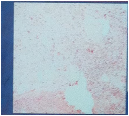
The tumor is histopathologically characterized by intense proliferation of myoblastic spindle cells wiyh chronic lymphocytic infiltration of the stratum.1,2 Three histological types are reported: a) mucosal, vascular and inflammatory areas resembling computed nodular peritonitis (Nodular fasciitis – like type), b) spindle cells intercalated with inflammatory cells (Lymphocytes, Eosinophils and Plasmacytes) resembling fibrous tissue (Fibrous histiocytoma - like type) and c) dense, squamous collagen, looks like fibrous scar tissue (Desmoid or scar tissue type) (Figure 3 & 4).

Figure 3 Histopathology specimen (HE, X100) with squamous cells, mucoid and inflammatory infiltration and spindle cells.
The tumor resembles laryngeal papillomatosis,13 squamous cell carcinoma, leiomyosarcoma, malignant histiocytoma, embryonic rhabdomyosarcoma and lymphoma and requires complete immunohistichemical tests (Vimentin, Muscle specific actin (SMA), ALK-1, Desmin, Caldesmon, Calponin, Cytokeratin, CD 68).14 Genetic rearrangements involving the 2p23 chromosome in which the ALK18 receptor tyrosine kinase gene has been mapped have also been observed.7,15
The diagnosis is based on histological examinations and is documented by immunodiagnosis and immunohistochemistry.1,2,3,16 In our case, the cells were positive for SMA, Caldesmon and Calponin while exhibiting inflammatory infiltration, myxoid degeneration and scattered spindle cells as in the second histological type above described.
Wening et al.,10 performed a review of 8 cases of inflammatory Myofibroblastic laryngeal tumor. The female to male ratio was 5 to 3, ages 19 to 69years old and duration of the symptoms from 10days to 4months. In the 7 patients the tumor was completely removed and the patients were free symptoms for up to 36months. One patient required surgery again after 2years and eventually after a few months underwent total laryngectomy.
VÖlker et al.,17 styding the international literature found that tumor appears more often and at older ages with an average age of 52years. The male-female ratio was 1.8 t0 1. They also found that the relapse rate was 21% and most of the cases in the first year after surgery.18‒21
In patients with chronic hoarsness who have a malignant - looking laryngeal tumor inflammatory myofibroblastic tumor should be considered. Conservatory surgery with occasionally additional steroid treatment and close follow up of the patient is recommended.
There is no exists any financial interest or any conflict of interest.
None.

©2017 Nikolaou,, et al. This is an open access article distributed under the terms of the, which permits unrestricted use, distribution, and build upon your work non-commercially.