Journal of
eISSN: 2379-6359


Case Report Volume 8 Issue 3
Department of Paediatic Surgery Pandit Bhagwat Dayal Sharma Post Graduate Institute of Medical Sciences, India
Correspondence: Jitu Sam George Department of Paediatic Surgery Pt B D Sharma Post Graduate Institute of medical science room no 180 doctors hostel Harayana124001, India, Tel 9496326191
Received: April 26, 2017 | Published: August 16, 2017
Citation: Rattan KN, George JS, Swati, Aman (2017) Infected Branchial Fistula Mimicking As a Branchial Cyst: Two Case Reports. J Otolaryngol ENT Res 8(3): 00245. DOI: 10.15406/joentr.2017.08.00245
Anomalies of the branchial arches are the second most common congenital lesions of the head and neck in children with second arch anomalies the commonest. Eventhough diagnosis of branchial arch anomalies are straightforward, sometimes atypical presentations may cause a dilemma in diagnosis. We are reporting two unusual cases of right infected branchial fistula mimicking as a branchial cyst.
Anomalies of the branchial arches are the second most common congenital lesions of the head and neck in children with second arch anomalies the commonest. Eventhough diagnosis of branchial arch anomalies are straightforward, sometimes atypical presentations may cause a dilemma in diagnosis. We are reporting two unusual cases of right infected branchial fistula mimicking as a branchial cyst.
Case 1
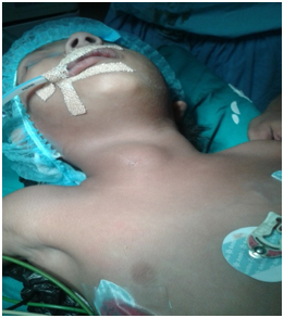
A six year old female child was brought to the paediatric surgery OPD of PGIMS, Rohtak with a complaint of swelling on right side of neck since birth associated with intermittent mucoid discharge. Swelling gradually increased in size for last one month with purulent discharge from pinhead sized opening over swelling at the junction of middle and lower one third of sternocleidomastoid. Swelling was cystic and non-tender with no overlying skin changes. A clinical diagnosis of infected branchial fistula was made on basis of clinical examination (Figure 1).

Figure 1 Case 1:- Preoperative picture showing swelling at the anterior border of right sternocleidomastoid.
CECT neck was done which confirmed the diagnosis. The patient was prepared for surgery under general anesthesia. An elliptical incision was given around the opening and flaps were raised. Around 20-30ml pus was drained. The tract was traced and found passing between internal and external carotid arteries and was opening into tonsillar fossa. Fistula was excised completely and sent for histopathology examination. Post-operative period was uneventful and wound was healthy. Excision of fistulous tract (Figure 2 & 3).
Histopathology revealed keratinized squamous epithelium lined tract with mucus secreting glands.
Case 2
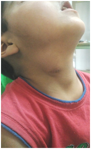
5year old child was brought to the paediatic surgery OPD of PGIMS Rohtak with complaints of right sided neck swelling of 4year duration which also increased in size over two months duration. On examination there was a swelling on the right side of the neck with an opening at its summit. Patient was admitted and was investigated. Imaging studies were done which confirmed the diagnosis of branchial fistula (Figure 4).

Figure 4 Case 2:- Preoperative picture with swelling in the anterior aspect of right sternocleidomastoid with an opening at its summit.
Excision was done under general anaesthesia. Initially pus was drained and the entire fistula tract was dissected and separated from surrounding tissue after giving an elliptical incision. On dissecting, the tract was found to be densely adhered to the carotid sheath. In this case also postoperative period was uneventful.
Developmental anomalies arising from branchial apparatus include cysts, internal sinuses, external sinuses and complete fistulas. Cyst may exist independently or anywhere along the course of a sinus or fistula. Most common clinical presentation in patients with branchial cysts consists of a left sided painless cervical mass. Patients with a branchial fistula usually presents with persistent mucus discharge from a skin opening in the neck usually in the second decade of life.2 The external opening is seen between the upper two thirds and the lower one thirds of sternocleidomastoid while internal opening is usually seen in the lateral wall of the pharynx region of tonsillar fossa. They are usually unilateral, commonly on the right side and are more prevalent in women even though bilateral cases are also reported.3,4 In the index cases patients presented with a right sided swelling with a narrow opening at its summit raising doubts that it can be a branchial fistula mimicking as branchial cyst.
Diagnosis of branchial anomaly is usually straightforward. However atypical lesions can be misdiagnosed. Correct diagnosis is very crucial as recurrence rates after surgical excision of branchial anomalies are 14% and 22% with previous infection and surgery, respectively, whereas the recurrence rate for primary lesion is 3%.5,6 Dench JE et al.,1 have reported a case in which a complete branchial fistula was mistaken for a right sided sebaceous cyst. Another case in which a low lying thyroglossal duct cyst with recurrent lateral cervical discharge and no palpable mass which was initially diagnosed as a second branchial arch fistula was reported by Sung-Min Hong et al.,7 In both these instances imaging studies helped in reaching the correct diagnosis. Fistulogram is one of the routinely performed and very useful investigations in the diagnosis of branchial fistula, in which the completeness of a fistula is diagnosed by a dye test in which methylene blue is injected through the outer opening and appears in the throat.8 A negative result can become positive during the time of general anesthesia due to muscle relaxation. Occasionally, the fistula tract may be blocked by secretion or granulation giving negative fistula test.5,9 However Rattan et al.,10 have reported in their study that fistulogram and dye injection was of no help during excision. Computed tomographic scanning (CT) and magnetic resonance imaging (MRI) of the neck are useful in delineating the branchial lesion and its relationship to surrounding neurovascular structures before any surgical intervention.9 It also helps in classifying the type of lesion, provides a roadmap for surgeon prior to surgery and reduces the chance of recurrence.
Treatment of choice for branchial fistula is complete surgical excision of the fistulous tract. Stepladder method and the stripping method is the two most commonly used surgical methods.3
So all cases with branchial arch anomalies should be thoroughly examined and properly investigated. Fistulograms can sometimes give false negative results but it should be complemented with CT and MRI which are almost always suggestive. Accurate diagnosis is an essential part of the management of second branchial arch anomalies which helps in reducing the chance of recurrence. A case of branchial fistula should be diagnosed and managed as early as possible as any delay can lead to secondary infection which can result in formation of adhesions with surrounding vital structures which might lead to complications during surgery.
None.
Author declares there are no conflicts of interest.
None.

©2017 Rattan, et al. This is an open access article distributed under the terms of the, which permits unrestricted use, distribution, and build upon your work non-commercially.