Journal of
eISSN: 2379-6359


Case Report Volume 15 Issue 1
1ENT and Head and Neck Surgery Department, Conakry University Hospital, Republic of Guinea
2ENT and Head and Neck Surgery Department, Republic of Guinea
3ENT and Head and Neck Surgery Department, Mamou Regional Hospital, Republic of Guinea
4ENT and Head and Neck Surgery Department, Labe Regional Hospital, Republic of Guinea
Correspondence: Keïta Abdoulaye, ENT and HNS Surgeon (Associate Professor), Conakry, Republic of Guinea, Tel (+224) 622442271
Received: January 25, 2023 | Published: February 27, 2023
Citation: Ibrahima D, Alseny C, Aliou DM, et al. Giant osteoma of the maxillary sinus: a case report. J Otolaryngol ENT Res. 2023;15(1):30-32 DOI: 10.15406/joentr.2023.15.00523
Objective: To report the management of a case of giant osteoma of the right maxillary sinus in a sub-Saharan ENT department.
Observation: The patient is 26 years old, housewife and without any particular history. She was referred to us by the oncology department for a right suborbital mass. The history goes back 4 years, with the appearance of a mass under the right orbit which has progressively increased in volume. She consulted at the indigeneity. Due to the persistence of the mass, she consulted an oncology department where the diagnosis of nasosinus lymphoma was made. She underwent 4 courses of chemotherapy. Without a favorable outcome, an ENT opinion was requested. The examination on admission noted a good general condition with stable hemodynamic constants. In the ENT sphere, there was a mass under the right orbit measuring 13x12 cm, painless, hard, not very smooth surface, regular edge, blackish skin opposite, with a mass effect in the homolateral nasal fossa and on the hard palate. The CT scan of the facial mass revealed a bone-toned hyperdensity at the expense of the right maxillary sinus isolated with extension to the adjacent soft tissues of the face and expression in the ipsilateral nasal fossa almost filling the maxillary sinus. Surgical excision via the paralateronasal approach of Moure and Sibileau was complete. The postoperative course was simple. Histology revealed a cancellous osteoma.
Conclusion: Osteoma of the maxillary sinus is less frequent in our practice. Its diagnosis is confirmed by histology. It has a good prognosis because of its benignity. Thus, it is important to be vigilant because it can be confused with malignant nasosinus tumors.
Keywords: osteoma, maxillary sinus, management
Osteomas of the maxillary sinus are rare benign tumors of slow evolution, most often asymptomatic, rarely developed at the expense of the maxillary sinus, whose natural history is poorly understood.1,2 The incidence of osteomas of the facial sinuses in the general population may be as high as 3%.3 Maxillary sinus osteomas account for only 14% of cases and rarely cause complications, but may occur as part of a syndrome.4,5 The treatment is surgical. The indication depends on functional signs and tumor size. Their recurrence is rare after adequate surgical excision.4
We report the management of a case of giant osteoma of the maxillary sinus at the ENT Department of the Donka National Hospital, Republic of Guinea.
The patient was 26 years old, housewife and without any particular history. She was referred to us by the oncology department for a right suborbital mass. The history goes back 4 years, with the appearance of a mass under the right orbit which progressively increased in volume. She consulted at the indigeneity. Due to the persistence of the mass, she consulted an oncology department where the diagnosis of nasosinus lymphoma was made. She underwent 4 courses of chemotherapy. Without a favorable outcome, an ENT opinion was requested.
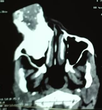
The examination on admission noted a good general condition with stable hemodynamic constants. In the ENT sphere, there was a mass under the right orbit (Figures 1a&1b) measuring 13x12 cm, painless, hard, with a smooth surface, regular border, blackish skin, with a mass effect in the homolateral nasal fossa and on the hard palate. The lymph node areas were free. The CT scan of the facial mass (Figure 2) showed a bone-toned hyperdensity at the expense of the right maxillary sinus isolated with extension to the adjacent soft tissues of the face and expression in the ipsilateral nasal fossa almost filling the maxillary sinus.

Figure 2 CT scan of the facial mass showing hyperdensity of bone tone at the expense of the isolated right maxillary sinus.
Surgical exploration by the paralateronasal approach of Moure and Sibileau (Figure 3) noted a bony and spongy mass at the expense of the right maxillary sinus with a mass effect in the homolateral nasal fossa. Complete removal was done by trepanation followed by milling (with round tungsten and diamond surgical burs). We did not record any intraoperative incidents or accidents. The postoperative care was marked by the administration of an antibiotic, analgesic and nasopharyngeal lavage. The postoperative course was simple. The patient was discharged on the 3rd day of her hospitalization. She was reviewed at one year without recurrence (Figure 4). Histology revealed a cancellous osteoma.
Sinus osteomas are benign tumors with mostly discrete symptoms and slow growth.3,5 They occur from the second decade onwards, as in our patient's case.6 The sex ratio varies according to the series.4,7 The etiopathogeny of osteoma is still obscure, hypotheses have been evoked, notably embryonic, traumatic and infectious.4,8 Pure frontal and fronto-ethmoidal osteoma represent 50 to 90% of all nasosinusal osteomas followed by ethmoidal osteoma (2 to 40%), the maxillary and sphenoidal forms are rarer.9–11 Orbital, neurological and endocranial extensions are the prerogative of ethmoidal or ethmoidofrontal forms.2 They have not been reported in our case.
The CT scan is the examination of choice. The osteoma presents as a single mass, with a sharp edge, spontaneously hyperdense, which does not enhance after injection of contrast medium.12 The histology is heterogeneous with ivory osteomas, formed of compact bone, and cancellous osteoma, comprising fibrous and medullary trabeculae.3
The approach in case of maxillary localization depends on the tumor size and the practitioner's habits: endoscopic or conventional surgery. In our patient we opted for the classical approach because of the large volume of the osteoma.5,13 The evolution is usually benign. Regular clinical and radiological follow-up is necessary because of the risk of recurrence, even late.2 In fact, she did not present any recurrence after 1 year.
Osteoma of the maxillary sinus is less frequent, occurring in the second decade of life. It may extend to the other sinuses, to the orbit and to the endocrine. The CT scan plays an important role in the diagnosis and the treatment remains surgical. However, we draw the attention of medical colleagues, especially oncologists, to consider this diagnosis in front of a hard and painless nasosinus mass.
None.
Authors declare no conflict of interests.
None.

©2023 Ibrahima, et al. This is an open access article distributed under the terms of the, which permits unrestricted use, distribution, and build upon your work non-commercially.