Journal of
eISSN: 2379-6359


Letter to Editor Volume 4 Issue 3
1Department of Otorhinolaryngology, La Milagrosa Hospital, Autonomous University of Madrid, Spain
2Department of Otorhinolaryngology, Puerta de Hierro-Majadahonda University Hospital, Autonomous University of Madrid, Spain
Correspondence: Pinilla Urraca M, Department of Otorhinolaryngology Head & Neck Surgery, C/ Joaquin Rodrigo, 228002 Majadahonda, Madrid, Spain, Tel +34 629810134
Received: June 07, 2016 | Published: July 5, 2016
Citation: Villarreal Patino IM, Pinilla Urraca M (2016) Dysphagia and Cervical Vertebral Osteophytes: Forestier's Disease? J Otolaryngology ENT Res 4(3): 00103. DOI: 10.15406/joentr.2016.04.00103
dysphagia, cervical osteophytes, forestier´s disease
DISH, diffuse idiopathic skeletal hyperostosis; DM, diabetes mellitus; MRI, magnetic resonance imaging
Cervical spinal osteophytes are estimated to affect 10 to 30% of the general population; nevertheless they tend to be largely asymptomatic. Symptoms as dysphagia, dysphonia and/or dyspnea can be observed when hyperostosis involves the anterior margin of the cervical vertebrae, more frequently (75%) in the population above 60years of age.1‒3
These skeletal abnormalities are considered by various authors as incidental radiographic findings rather than a disease. However, in these cases, an entity to consider is a systemic rheumatological abnormality of unknown etiology, diffuse idiopathic skeletal hyperostosis (DISH) or Forestier´s disease described in 1950.1 It produces calcification-ossification of the anterior longitudinal ligament. The low dorsal region is the most affected in the spine.4 Dysphagia is the most common symptom when the disease affects the cervical spine (C3-C5 levels), less frequent is dyspnea, both secondary to extrinsic compression of the esophagus and trachea. Forestier’s disease is not rare, but it is often undiagnosed. Nowadays, the reported prevalence of DISH ranges from 3 to 30%, it affects men more frequently than women (2:1), the peak occurrence is in patients in their 60s.2,5
Dysphagia in these cases typically begins with as discomfort when swallowing solids and progressing to liquids, progressively evolving toward dysphagia. It is commonly progressive in nature and acute presentation is very unusual. There are several theories to explain dysphagia:2,5‒7
Numerous etiological hypotheses have been made regarding risk factors associated with DISH such as trauma, vitamin A exposure, and endocrine and metabolic factors such as obesity, type 2 diabetes mellitus (DM), hyperuricemia, and dyslipidemia. Nevertheless, definitive associations have yet to be studied and developed. There is no conclusive correlation between symptoms and the size of the osteophytes: however, advanced age has been correlated with the severity of symptoms. This leads to various reports finding the strongest correlation with DISH being sex and age.1,2,5,6
Denko and Malemud found that DISH patients tend to have higher insulin and growth hormone levels.2,3 Growth hormone induces local production of insulin-like growth factor-1, which in turn stimulates alkaline phosphatase and type-2 collagen activity in osteoblasts.
A recent study suggested that alterations in gene expression might play a role in this type of hyperostosis (including collagen synthesis involvement). Recently, low levels of DKK-1 (involved in regulating bone formation and regeneration) have been linked to cases of severe ossification.31 this finding concludes that an alteration in DKK-1 may contribute to skeletal hyperostosis.1,2 Radiological imaging is undoubtedly indispensable for DISH to be diagnosed.2,5 They include X-ray, CT, and/or magnetic resonance imaging.
The specific radiological criteria for the diagnosis of Forestier’s disease were described by Resnick and Niwayama in 1970 and are still used today:1‒3,5,6,8
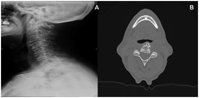
A characteristic radiographic feature of spine involvement is the presence of hyperostosis of the cortex along the anterior surface of the vertebrae (Figure 1). Gradually, elongated Osteophytes occur at the anterior margin of the vertebrae and grow across the disc space. Dysphagia from cervical osteophytes (usually located in the anterior and lateral regions of the vertebral bodies) has ranged from C3 to C6. Osteophytes originating from the level of the fifth and sixth cervical vertebrae are usually the ones implicated in this symptom.2

Figure 1A: Lateral cervical X-Ray image B: CT axial view; both showing hyperostosis of the cortex along the anterior surface of the vertebrae. Gradually, elongated osteophytes occur at the anterior margin of the vertebrae and grow across the disc space.
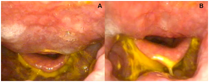
Some authors perform magnetic resonance imaging (MRI) looking moreover for intramedullary involvement. Potential mechanical obstruction might be assessed with nasal endoscopy (Figure 2), barium swallow, esophagram, and video fluoroscopic studies.8

Figure 2A, B Endoscopic view during a dysphagia clinical assessment of a patient with Forestier’s disease. Notice the minimum space between the epiglottis and the posterior pharyngeal wall
Other causes of dysphagia should discard before retaining osteophytic compression as imputable. Ankylosing Spondylitis, spondylosis deformans, degenerative diseases of the intervertebral discs, osteoarthritis have to be ruled out.5,8 Symptoms and radiological signs of DISH can be mimicked by these and other less common causes such as disorders of esophageal mobility, vascular abnormalities, and neurological disorders. Also the possibility of a malignancy should not be overlooked.
The treatment of DISH is based on a conservative multidisciplinary approach.2 Conservative treatment seems plausible to have an effect on reducing inflammation and swelling (at least temporarily). Dysphagia may be treated to begin with nonsurgically with anti-inflammatory medication (including steroids), antireflux medication muscle relaxants, dietary restrictions, speech and swallow therapy. Postural changes when swallowing may also be necessary. These methods may offer temporary relief in patients affected by dysphagia, long-term treatment often is difficult due to its poorly understood etiology. Medications including biphosphonates have also been proposed as part of Forestier´s treatment.1,9,10
Indications for surgical treatment are mainly after cervical trauma, airway impairment, and dysphagia refractory to conservative treatment. It may also be envisioned when dysphagia is so severe and approaches aphagia or if the patient has experienced substantial weight loss. Surgical treatment for DISH involves a simple osteophyte excision (osteophytectomy) to begin with. Clinical improvement might be achieved only with partial resection, or even resection of only the largest osteophytes taking into consideration that complete resection is not always possible.2,5,11
Surgical procedures include the anterolateral, posterolateral, or transoral approaches. The anterolateral approach (Smith-Robinson approach) is preferred for lesions affecting the lower level of the cervical spine. This access provides a rapid and good exposure to the great vessels and neurological structures. The posterolateral access involves a higher risk of injury to the sympathetic chain. It is usually used to approach C3-C6. The DISH mainly affects the C3-C6 region the antero-lateral neck approach is considered by most of the authors. An aesthetic advantage and a lower risk of affecting neurovascular structures can be achieved via the transoral or transpharyngeal approach. High cervical column lesions (C1-C2) may require this via.5,7‒11 Additional surgical procedures might be required, including spinal decompression, discectomy, spinal fusion, and secondary tracheostomy.10,11
Postoperative dysphagia can also be observed due to damage to surrounding structures, most significantly damage to recurrent and superior laryngeal nerves. Other postreatment complications include pneumonia; neuropraxia of laryngeal nerve, neuropraxia of the marginal mandibular nerve, esophageal motility disorders, stroke and death.8,10‒12
Recent studies indicate that ossifications may reoccur several years after successful initial surgical resection.10 Dysphagia due to cervical involvement of Forestier’s disease is a rare entity. Complementary exams are indispensable to make a positive and differential diagnosis as well as their therapeutic possibilities. Factors such as obesity, diabetes mellitus, and a metabolic syndrome are all positively associated with the development and progression of the disease. Definitive natural history and diagnostic criteria remain controversial.
None.
Author declareas there are no conflicts of interest.
None.

©2016 Villarreal, et al. This is an open access article distributed under the terms of the, which permits unrestricted use, distribution, and build upon your work non-commercially.