Journal of
eISSN: 2379-6359


Salivary gland tumors are usually rare constituting 3% to 4% of all head and neck neoplasms. Lymphoepithelial carcinoma, a type of squamous cell carcinoma, is a poorly differentiated carcinoma characterized by infiltrative lymphoplasmocytic aspect. It represents 0.8% to 2% of oropharyngeal malignancies. Most of these tumors are specifically found in the tosnils and base of the tongue, but can also be found in other adjacent structures.1
Keywords: salivary gland, nasopharyngeal carcinoma, lymphoepithelial carcinomas, Epstein Bar Virus, malignant neck tumors
Lymphoepithelial carcinoma was first described in 1921 by Regaud and Schminke as a tumor of the nasopharynx, an undifferentiated carcinoma with predominant lymphocytic infiltration.2 Lymphoepithelial carcinoma is a very rare type of nasopharyngeal tumors which can also affect salivary glands and the parotid gland specifically.3 A relatively small number of cases have been reported since the first encounter with this tumor in the salivary glands, being less than 150 cases in a 40 year period.4 Lymphoepithelial carcinoma can be commonly found in organs other than salivary glands such as lungs, ovaries, and breast.
These findings were related to specific geographical regions as they were diagnosed in Chinese and Eskimo populations.2 In these populations, the tumor mostly involves the parotid gland with a greater incidence is found in females. Some recent research found the greater incidence of patients with this tumor to be in Caucasians in the age group 50-70, but with no gender preference.3
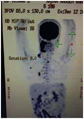
A 60 year old female was presented to the Otolaryngology Department at Hammoud Hospital University Medical Center for right neck mass. The patient was known to have diabetes type 2, hypertension and coronary artery disease status post open heart surgery. The mass was at the site of the parotid gland, painless, firm, rubbery, and non-mobile. Computerized tomography scan (CT scan) of the neck with intravenous contrast was ordered and done. It revealed presence of enlarged right parotid gland with irregular borders and locally enlarged lymph nodes suggestive of malignancy (Figure 1). A total body positron emission topography scan was done afterwards and revealed a malignant picture of the right parotid gland and the surrounding lymphatic structures. Right total parotidectomy with lymph node dissection was scheduled (Figure 2).

Figure 2 PET scan of our patient showing primary malignant salivary gland tumor with local and distant metastasis.
Under general anaesthesia, a modified Blair incision was done on the right aspect of the neck, followed by dissection and excision of both lobes of parotid gland, superficial and deep, with preservation of the facial nerve and its branches. Caution was taken to preserve nearby vessels and structures. Further dissection was done and four lymph nodes were excised from the right sub-mandibular and right posterior triangles and sent to pathology. Proper haemostasis was done and the incision sutured after placing a temporary drain.
The pathology report revealed presence of lymphoepithelial carcinoma of the right parotid gland with positive lymph nodes from specimens taken. Upon performing pathological studies, the sample excised was stained with hematoxylin and eosin stain (Figure 3) which showed lymphocytic infiltrates invading the parotid gland, EBER stain (Figure 4) showed positive results for presence of Epstein bar virus. The excised lymph nodes were also stained (Figure 5), and metastasis was found.
Lymphoepithelial carcinoma is a rare tumor of the salivary gland. It can be found in multiple other parts of soft tissues.2 It was first described by Hilderman in 1962 as a type of malignant salivary gland tumor which comprises 0.3-5.9% of these tumors.5 Upon histopathologic examination, it was found to have similar characteristics to non-keratinizing undifferentiated nasopharyngeal carcinomas. The immunohistochemical staining showed a similar pattern to NPC (Nasopharyngeal Carcinoma), as tumor cells were always positive for AE1/AE3, CK5/6 and p63 (Figure 6).3 It was reported to be in close contact with EBV in some cases(2) and to both Human Papilloma virus and EBV in other patients.2

Figure 6 Positive Immunostaining of PLEC, AE1 (A), cytokeratin (B), EMA (C), p63 (D).3
This tumor has a higher incidence in Eskimo and Chinese population in ages between 30 and 80 years with a male predominance.2 However, other recent studies showed a greater incidence in Caucasians in the middle age group with no gender predominance,3 which raises questions about the true origins and patterns of this rare malignant tumor.
Patients often present with painless slowly growing palpable masses. Computerized tomography scan and magnetic resonance imaging are done as means of diagnosis, but these do not allow discrimination from other salivary gland malignant tumors.5 Upon histological examination, the specimens show well circumscribed nodules characterized by nests with prominent lymphoid stroma and features of squamous cell carcinoma.5
These are differentiated from nasopharyngeal carcinoma by endoscopy and CT scan with intravenous contrast (Figure 7).5

Figure 7 Axial contrast enhanced CT, and axial gadolinium enhanced T1 weighted imaging of malignant salivary gland tumor.5
Lymphoepithelial carcinoma is a type of malignant poorly differentiated salivary gland tumor of rare incidence. This tumor usually occurs in sites other than salivary glands. The main method for treatment is surgical excision followed by radiotherapy, as this was the most successful method since this type was first identified in 1962.
None.
The author declares there are no conflicts of interest.

© . This is an open access article distributed under the terms of the, which permits unrestricted use, distribution, and build upon your work non-commercially.