Submit manuscript...
Journal of
eISSN: 2379-6359

Otolaryngology-ENT Research

Correspondence:

Received: January 01, 1970 | Published: ,

Citation: DOI:

Download PDF

Keywords

otitis media, clinical entity, extra cranial and intracranial complications, delta-sign, thrombus

Abbreviations

LST, late stent thrombosis; MRI, magnetic resonance imaging; CT, computed tomography

Introduction

Lateral sinus thrombosis (LST) is a major intracranial complication of otitis media. In preantibiotic era, prognosis was bad and it usually occurred in association with other intracranial complications.1,2 LST occurs by following mechanisms:

  1. by thrombophlebitis of the venules surrounding the external dural sinus wall
  2. by erosion of the bone over the sigmoid sinus by cholesteatoma
  3. by the infection that spreads to the sigmoid sinus through a bony dehiscence

It usually occurs as a complication of attico antral type of chronic otitis media and here direct dissemination of the infection will occur through the adjacent eroded bone. Many authors have reported lateral sinus thrombosis in patients with intact bony sinus plate. This suggests thrombophlebitic spread through the small emissary vein.3‒7 Its incidence LST is decreased because of the availability of good broad-spectrum antibiotics, availability of CT and MRI scans and micro surgical treatment. Now LST is a rare complication of otitis media. Otologist should be familiar with this clinical entity and it should be diagnosed early for good outcome.1,8 Radiological investigations play an important role in the diagnosis of LST. Definitive diagnosis of LST is made at surgery. CT and MRI are the investigations that are needed for correct diagnosis. In LST, CT scan with contrast shows a classic ‘delta sign’ of perisinus dural enhancement and filling defect of the lateral sinus (Figure 1). The ‘delta sign’ is not always detectable in CT.I patients with LST, along with MRI, CT scan also should be done to rule out other associated extra cranial and intracranial complications of otitis media.9‒14

MRI is more accurate than CT in detecting the thrombus. It demonstrates blood flow and accurately shows the site of sinus obstruction (Figure 2). It also shows subsequent reversal of flow. On MRI with contrast (gadolinium), thrombus appears as soft tissue signal associated with vascular bright appearance of the dural wall. This is known as the “delta-sign” (Figure 3).10,11,15,16 In addition, MR venography will also show the loss of signal and the absence of flow in the sinus (Figure 4). It has proven to be a valuable diagnostic tool in identifying LST.13 In cases of LST, MRI is the investigation of choice. It should be done in along with CT, to rule out any associated otologic and intracranial complication.16,17
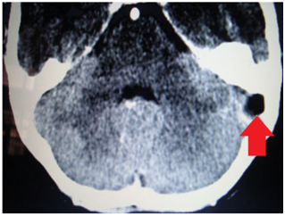
Figure 1 Light miscroscopyunder transmitted light –Showed enamel rods in light brown colour. Enamel rods showed alternate T.S and L.S of hunter Schreger bands. Histology of enamel under light microscopy showing alternate L.S (white arrow) and T.S (yellow arrow) of enamel rods (transmitted light).

Figure 2 CT with contrast showing (arrow) absence of enhancement in the thrombosed left lateral sinus and enhancement of the dura surrounding the sinus (delta sign).

Figure 3 MRI showing (arrow) occlusion and dilatation of left lateral sinus.

Figure 4 MRI venography showing (arrow) absence of flow in the right lateral sinus.

Acknowledgements

None.

Conflict of interest

Author declares that there is no conflict of interest.

References

  1. Viswanatha B. Lateral sinus thrombosis in children: A review. Ear Nose Throat J. 2011;90(6):E28‒E33.
  2. Viswanatha B, Naseeruddin K. Complications of atticoantral otitis media Revisited. Advances in Research. 2014:2(11);666–695.
  3. Tveteras K, Kristensen S, Dommerby H. Septic cavernous and lateral sinus thrombosis. Modern diagnostic and therapeutic principles. J Laryngol Otol. 1988;102(10):877‒882.
  4. Hawkins DB. Lateral sinus thrombosis: A sometimes unexpected diagnosis. Laryngoscope. 1985;95(6):674‒677.
  5. Murphy TP, Boydston W. Lateral sinus thrombosis. Otolaryngol Head Neck Surg. 1997;117(6):S134‒S137.
  6. O’Connell JE. Lateral sinus thrombosis: A problem still with us. J Laryngol Otol. 1990;104(12):949‒951.
  7. Viswanatha B, Naseerudin K. Lateral sinus thrombosis in otology: A review. Mediterranean Journal of Hematology and Infectious Diseases. 2010;2(3):e2010027.
  8. Amirmajdi NM. Sigmoid sinus involvement in middle ear infection. Laryngoscope. 1988;98(3):310‒312.
  9. Viswanatha B. Lateral sinus thrombosis: A major problem still with us. Indian journal of Otolaryngology & head and neck surgery. 2007;59(1):24‒27.
  10. Kaplan DM, Kraus M, Puterman M. Otogenic lateral sinus thrombosis in children. International journal of pediatric Otorhinolaryngology. 1999;49:177‒183.
  11. Levine SC, DeSouza SC. Intra cranial complication of otitis media. In: Glasscock III ME, Gulya CJ, editors. Shambaug’s Surgery of the Ear. 5th ed. Ontario: BC deckers Inc; 2003. p. 443‒461.
  12. Leiberman A, Lupu L, Landsberg R, et al. Unusual complications of otitis media. Am J Otol. 1994;15:444‒448.
  13. Irving RM, Jones NS, Hall-Craggs MAH, et al. CT and MRI imaging in lateral sinus thrombosis. The Journal of Laryngology and Otology. 1991;105(8):693‒695.
  14. Viswanatha B. Lateral sinus thrombosis with cranial nerves palsies. International Journal of Pediatric Otorhinolaryngology extra. 2007;2(3):165‒168.
  15. Dubey SP, Larawin V. Complications of chronic suppurative otitis media and their management. Laryngoscope. 2007;117(2):264‒267.
  16. Irving RM, Jones NS, Hall-Craggs MAH, et al. CT and MRI imaging in lateral sinus thrombosis. The Journal of Laryngology and Otology. 1991;105:693‒695.
  17. Kale US, Wight RG. Primary presentation of spontaneous jugular vein thrombosis to the Otolaryngologist- in three different pathologies. The journal of Laryngology and otology. 1998;112(9):888‒890.
Creative Commons Attribution License

© . This is an open access article distributed under the terms of the, which permits unrestricted use, distribution, and build upon your work non-commercially.