Journal of
eISSN: 2379-6359


Cystic masses of neck consist of various pathologic entities. Epidermoid cysts are rare in the head and neck region. Patient presents with a painless slow growing swelling. Imaging modality used to diagnose and manage the epidermoid cyst is ultrasonography followed by CT scan. In this study, diagnosis and management of a giant epidermoid cyst in the submental region is being considered.
Keywords: submandibular region, epidermoid cyst, excision
Epidermoid cysts are cystic swellings occurring in patients between 2nd to 4th decades but can manifest in any age group. These are usually asymptomatic but they can cause dysphagia, dysphonia and dyspnea once increases in size.1 There are various studiesreportingpain, speech disorder, or respiratory distress due to epidermoid cyst in the oral cavity, lower lip, or upper lip.2 Giant epidermoid cysts are rare and they usually present in the scalp.
Epidermoid cysts are relatively less common in the head and neck region, hence are likely to be misdiagnosed.
A 20 year old female patient came to ENT out-patient department with chief complaint of a painless and progressively increasing mid line neck swelling in the submental region of 5 to 6 years duration and simultaneous swelling in the floor of mouth (Figure 1). She also had a history of difficulty in speaking. On examination, a cystic, mobile, non-tender and non trans-illuminant swelling of 8cm x 8cm was observed in the submental region extending almost from one angle of mandible to another. The swelling did not move on protrusion of the tongue or swallowing. The intraoral swelling was present at the floor of mouth, pushing the tongue upwards and causing difficulty in speaking. There were no other palpable neck masses or enlarged lymph nodes. Routine haematological and the biochemical investigations were normal.
On Ultrasound study, a well-defined, unilocular, thick walled, round shaped, cystic swelling in the submental region, deep to mylohyoid muscle was found. Multiple, well defined, echogenic nodules, each measuring between 3 to 4 mm, were appreciated within the cystic lesion. After-shadowing or post acoustic enhancement from the nodules was not observed.
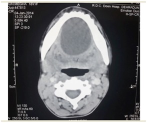
FNAC revealed epidermal inclusion cyst. Contrast enhanced CT imaging of the neck was done and it showed a mass measuring 8 × 8 cm with a clear boundary and regular margin positioned under the platysma anterior to the bilateral submandibular gland. Margin was enhanced in a ring pattern and the mass displaced the submandibular gland posteriorly (Figure 2).
The cyst was surgically excised completely by submental approach and excised mass was sent for histo-pathological examination (Figure 3). The intraoral component of the cystic swelling was removed by midline intra-oral incision. No intra or post operative complications were seen.
Specimen on gross examination after cutting was filled with cheesy white material. Histopathology revealed fibrocartilaginous cyst wall lined by stratified squamous epithelium with presence of granular layer. The cavity showed lamellated keratin and flakes. Adjacent tissue showed mild chronic inflammation.

Figure 2 Contrast enhanced CT imaging of the neck was done and it showed a mass measuring 8 × 8 cm with a clear boundary and regular margin positioned under the platysma anterior to the bilateral submandibular gland. Margin was enhanced in a ring pattern and mass displaced submandibular gland posteriorly.
The spectrum of swellings originating from a teratoma includes dermoid cyst, epidermoid cyst and teratoid cyst, all lined by squamous epithelium.3 Dermoid and epidermoid cysts are inclusion cysts lined with ectoderm having difference in the contents within. Dermoid and epidermoid cysts are unusual swellings in the head and neck region representing only 7 % of all cystic swelling in that region.4 All these cysts are lined by squamous epithelium and contain cheesy keratinaceous material.
Epidermoid cysts may be congenital or acquired in origin, but cannot be diffentiated histopathologically. Epidermoid cysts do not have dermal appendages and the squamous lining is thin and devoid of any calcifications. Due to continuous desquamation of the epithelium lining from the cysts, debris is formed consisting of keratin, proteinaceous material and some cholesterol. On gross examination, the lesions are called as ‘pearly’ due to the shiny smooth, waxy appearance of the dry keratin.5
The incidence of the dermoid and epidermoid cysts comprises 7 % of all head and neck cystic swellings. These swellings most commonly occur near the lateral eyebrow. Out of all the dermoid cystic swellings occuring in the head and neck region, 11.5 % are observed in the submental region 6. Epidermoid cysts, occurring particularly in the neck region, are less common than the dermoid cysts and they occur most commonly in the submental region.
These cystic swellings generally present as midline neck masses which gradually increase in size over the years due to deposition of cutaneous products. The lesions are soft in consistency, mobile and the overlying skin can be easily pinched. These swellings do not move with the protrusion of the tongue or the swallowing movement. These cysts can assume the size varying from few millimeters to 12.0 cms.7,8,9
On ultrasonography, the epidermoid cysts exhibit as thick walled cysts with echogenic debris containing within. Multiple well defined, dependent, echogenic nodules may be seen in the cysts. On computed tomography, the epidermoid cysts show low attenuation. On magnetic resonance imaging, the epidermoid cysts are hypo-intense on the T1 weighted images and hyper-intense on the T2 weighted images, with multiple hypointense nodules within.
The management strategy consists of complete surgical excision and simultaneously taking precautions to not rupture the cysts, as the ruptured substance may act as irritant to the surrounding fibrovascular tissues.10
The differential diagnosis of the sublingual lesions includes lymphatic malformations, dermoid and epidermoid cysts and enlarged lymph nodes. Imaging helps to a greater extent in diagnosing and disnguishing dermoid and epidermoid cysts. The prognosis is very good, with a low incidence of recurrence. The malignant transformation of these cysts has not been reported.
Epidermoid cysts of the submental region are slow growing swellings. Diagnosis is made by ultrasonography followed by CT Neck, confirmed by fine needle aspiration cytology. Dermoid and epidermoid cysts are rare in neck (7%). Treatment consists of complete surgical removal of the cyst.
None.
The author declares there is no conflict of interest.

© . This is an open access article distributed under the terms of the, which permits unrestricted use, distribution, and build upon your work non-commercially.