Journal of
eISSN: 2379-6359


Aim: Decannulation of tracheostomy in cases of traumatic quadriplegia is always a challenge due to respiratory muscle paralysis, chest infections, aspiration and need to care for sacral sores. Aim of this study was to identify and overcome the difficulties in decannulation of tracheostomy in cases of quadriplegia due to cervical spinal cord injury.
Material and methods: This prospective observational study was carried out in a tertiary care Spinal Cord Injury Center of a Military Hospital. Ten cases of tracheostomized traumatic quadriplegics were evaluated over 3 years. Decannulation was considered once there was normal gag reflex, effective cough, manageable aspiration, oxygen saturation above 90%, no serious pulmonary compromise and no need for bed sores surgery. The cases were followed up for further one year post-decannulation.
Results: Eight cases could be decannulated successfully. Only two cases could not be decannulated due to feeble chest movement, ineffective Cough, lack of motivation and occasional aspiration.
Conclusion: Cervical cord injury patients have difficult decannulation due to weak respiratory muscles, ineffective cough, aspiration, pulmonary pathology and bed sores. Respiratory muscle exercise, quantified peak cough pressure, assisted coughing, risk benefit approach in aspiration, healing of bed sores and team work are keys to successful decannulation.
Keywords: tracheostomy, decannulation, spinal cord injury, quadriplegia, respiratory muscle paralysis, peak cough flow rate
Decannulation in the process of taking out the tracheostomy tube permanently and closure of wound after the patient has settled down for the disease for which tracheostomy was performed.1 Although tracheostomy is considered to be the most common surgical procedure performed on critically ill patients, there is no general consensus as to when a tracheostomy tube can be safely removed.2 Commonly encountered difficulties during decannulation are tube dependency, bleeding, and mechanical problems with cuff, aspiration, pulmonary complications, tracheomalacia, scarring of the tracheostomy wound, inversion of wound margin in skin, tracheal stenosis and also migration of granulation tissue inside the trachea through the tracheostomy wound. Patients with traumatic quadriplegia following cervical spinal cord injury (CSCI), who cannot self care for their tracheostomy, pose more difficult situations due to paralysis of respiratory muscles, concomitant chest infections, problems of aspiration and the need to care for sacral bed sores. Decannulation in these cases is always a challenge. These patients are generally bed and wheel chair bound, in need of a constant attendant for daily chores and in a state of psychological trauma. In situ tracheostomy tube for prolonged period of time adds to the woes. Removal of tracheostomy tube make them able to communicate verbally, restores their self esteem by removing the dependency on suction apparatus and enabling oral feed and improves their health and quality of life. A prospective observational study was conducted in a tertiary care Spinal Cord Injury Centre (SCIC) of Armed Forces Medical Services of India to understand the decannulation problems in quadriplegic patients of CSCI and to study the causes of difficulties in decannulation in these cases.
33 cases of traumatic quadriplegia due to CSCI were treated in Neurosurgical centre and transferred to our SCIC between August 2012 and August 2014. 16 of them had undergone tracheostomy, out of which, six were already decannulated in neurosurgical centre. Remaining ten patients, with tracheostomy tube still in situ, were studied in our centre over two years for the difficulties encountered during the attempts made to decannulate them. Approval from ‘Hospital Ethical Committee for Clinical Research’ was obtained to carry out the study. Other spinal cord injury patients who had not undergone tracheostomy or who had already been decannulated in Neurosurgical centre were excluded from study. Cases of tracheostomy in patients other than spinal cord injury, such as head injury, were also not included in the study. There were nine male and one female patient in the age group of 13 years to 52 years. All had suffered trauma to different levels of cervical spinal cord by various causes. Medical details of the patients are as in Table 1. All had undergone neurosurgical procedures, had been on ventilatory support during post-operative phase and had undergone tracheostomy anticipating prolonged ventilatory support. None had head injury.
A detailed chart review was performed in each patient. Indication for tracheotomy was confirmed to have been resolved. The patients had already been weaned from ventilators. Airway patency was confirmed by fibreoptic laryngoscopy. Vocal cord function was assessed by voice quality during temporary occlusion of the tracheostomy tube with deflated cuff and also by Hopkin rod lens laryngoscopy. Vocal cords were normal, moving symmetrically and approximating in the midline on phonation. A downsized double lumen fenestrated tracheostomy tube was inserted in place of the original one for easy maintenance and to aid phonation. Tube was corked for 24 hours as trial.
Decannulation was, thereafter, considered if following criteria in our protocol were fulfilled:
Once the above criteria were fulfilled, tracheostomy tube was removed in the morning under close observation of the trained nurse. The nurse kept an hourly watch on the patient’s oxygen saturation, need for suction clearance of secretion, strength and effectiveness of cough and any distress of the patient. A spare tracheostomy tube of a size smaller, along with a tracheal dilator, was kept at bedside for emergency recannulation.
Total of 10 cases were studied over a period of 2 years from August 2012 to August 2014. These cases were further followed up for another year up to August 2015. 8 out of 10 patients (80%) were within 13 to 30 years of age. Table 1 show that majority of the cases (60%) were due to sports injuries, probably because our subjects were soldiers who actively pursue sports. Most of the cases (30%) suffered injury at C4 and C5 vertebral level. Majority of the cases (60%) had anterior subluxation of vertebra with cord contusion. There was only one case of complete cord transaction. Table 1 also shows that neurological recovery can be varied and unpredictable and may not be favoured by age or level of cord lesion.
Cases |
Age/Sex |
Nature of injury |
Cause |
Neurosurgery done |
Period of venti- |
Venti- |
Tracheo-stomy |
Associated problems developed in hospital |
Main issue for decannulation |
|
Case 1 |
13/F |
Fracture CV-4&5 with Cord contusion |
RTA |
Discectomy CV 4/5, plating & cage placement C 4 to 6 |
15 days |
1 days |
59 days |
Vesicular calculi |
Week cough |
|
Case 2 |
23/M |
Anterior subluxation C3-4 with cord compression |
Fall from pole |
Discectomy C3-4, iliac crest grafting & titanium plating |
10 days |
8 days |
126 days |
Sacral sore, |
Week cough |
|
Case 3 |
22/M |
Bilateral facetal dislocation, cord transaction C5/6 |
Wrestling |
Reduction, discect- omy, spacer, ant plating C 5/6 |
12 days |
4 days |
172 days |
Pneumothorax (right), Sacral sore |
Week cough |
|
Case 4 |
52/M |
Ant Subluxation C6/7 with cord contusion |
Fall from tree |
C3-5/D1-4 interspinous wiring with bone grafting |
06 days |
6 days |
5 years |
Sacral sore, Autonomic dysreflexia, Herpes zoster, Gastric ulcer |
Week cough Aspiration |
|
Case 5 |
40/M |
Anterolisthesis C4/5 |
Gymnastic |
Reduction and anterior plating |
4 days |
4 days |
86 days |
Sacral sore, |
Week cough |
|
Case 6 |
20/M |
Ant PIVD C4/5 disc bulge |
Judo |
Discectomy, Fusion C4/5 with plate screw & iliac crest |
6 days |
6 days |
44 days |
Nephrogenic diabete insipidus |
Week cough |
|
Case 7 |
23/M |
Anterolisthesis C5/6 , disc extrusion & epidural hematoma |
Wrestling |
Reduction, discectomy, fusion C 5/6 with plate & screw |
8 days |
8 days |
215 days |
Sacral sore, Pneumonia |
Week cough |
|
Case 8 |
19/M |
Anterior subluxation C2-3 with cord compression |
Diving |
Discectomy C2-3, |
8 days |
8 days |
710 days |
Sacral sores, Pneumonia, Depression |
Week cough |
|
Case 9 |
20/M |
Ant Subluxation CV-5 with Cord compression |
Fall of a hatch over head |
Anterior fixation of CV4 to CV6 |
37 days |
3 days |
41 days |
Collapse left lung, |
Week cough |
|
Case 10 |
30/M |
Fracture CV-4, 5 & 6 with Cord compression |
RTA |
Corpectomy C5 – C6 with anterior plating C4 to C7 |
11 days |
7 days |
3 years |
DVT left leg, Cardiac arrest, Sacral sore, |
Weak cough, |
|
Table 1 Medical details of the quadriplegic patients
Abbreviations: CV, cervical vertebra; RTA, road traffic accident; PIVD, prolapsed inter-vertebral disc; DVT, deep vein thrombosis; C, cervical; D, dorsal; F, female; M, male;
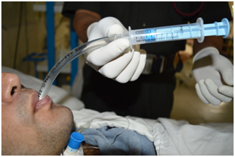
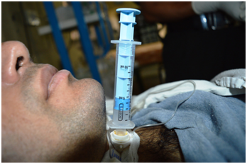
A case of single vertebral level involvement at C5 as also a case of prolapsed intervertebral disc which is known to have good surgical result could be decannulated early on 41st and 44th days respectively. A young patient of 19 years (Case 8), with high cord lesion at C2-C3, took 2 years and 10 days to decannulate whereas a 30 years old (Case 10), with mid-cord lesion at C4-C5-C6, is on tracheostomy even after 3 years and a case of low cord injury at C6-7 (Case 4) could not be decannulated even after 5 years 6 months. Factors found to be adversely affecting decannulation were respiratory muscle weakness, ineffective cough, associated pulmonary disease, other medical co-morbidities and the need for reconstructive surgery for sacral sores. Eight cases could be decannulated after observing the patients under our protocol mentioned above. Two cases, however, could not be decannulated. One of them (Case 4) continues to have feeble chest movement, ineffective cough and occasional aspiration and the other (Case 10) has ineffective cough and complete lack of motivation. His motivation level has not yet improved in spite of indoor support of buddy soldier as attendant, bed side care by his wife and periodic counselling by Psychiatrist. The difficulties observed are depicted in Table 2. Patients of CSCI often undergo tracheostomy if it is anticipated that they are going to require ventilator support for more than 3 weeks.3 The frequency of tracheostomy in patients with traumatic quadriplegia has been reported to range from 16% to 30% with a median of 31 days from the day of tracheostomy to that of decannulation.4 Tracheostomy tube in situ for a prolonged period poses numerous risks such as tube dependency, tracheal stenosis, trachea-cutaneous fistula, dysphagia, aspiration, stomal infection, pulmonary complications, accidental dislodgement etc. Tracheostomised patient are unable to vocalize unless they can tolerate cuff deflation. Quadriplegic patients are particularly disadvantaged by this because they are also unable to write. The inability to communicate effectively can be immensely distressing for the patient and their attendants.5 Literature shows a considerable diversity in criteria for decannulation. Stelfox and colleague, in their survey in 2009, observed considerable differences in the decannulation practices, which necessitated the need for development of evidence based trachesotomy decannulation guidelines. They identified clinician factor and patient factors as the two determinants of trachesotomy decannulation. Clinician factor was principle work facility whereas the patient factors included oxygenation, level of consciousness, ability to tolerate tracheostomy tube capping, effectiveness of cough and aspiration.6 However there are few specific considerations in CSCI patients which include neurological level of injury, completeness of cord injury, respiratory muscle weakness, vital capacity and peak cough flow, airway hyper-responsiveness, aspiration and care for bed sores. Respiratory dysfunction resulting from cervical spinal cord injury depends on the level of injury and the extent of denervation. Higher level lesions result in progressive denervation of expiratory and inspiratory muscles. Complete paralysis of all muscles involved with respiration occurs when the lesion is above C3. Lesion between C3 to C5 causes respiratory insufficiency due to paralysis of diaphragm. Although primary and some accessory muscles of inspiration remain fully innervated with injuries below cervical lesion, the ability to ventilate at higher levels or during exercise is still compromised because intercostals and other chest wall muscles do not provide integrated expansion of upper chest wall when the diaphragm descends during inspiration. Partially or fully denervated expiratory muscles in SCI patients also diminish exercise ventilation and ventilatory reserve.7 Although the primary consequence of cervical cord injury is denervation of the respiratory pump, secondary consequence occurs within the lungs because of the inability to effectively distend and inflate to its full capacity. The compliance of the lungs goes down with time. Inspiratory muscles are ideally trained using a resistive and threshold trainer device. It has a one way valve that closes during inspiration so that patient must breathe against a load.8 In place of this commercial device, weakness of the respiratory muscles was overcome in our institute by making the patients use simple hand held devices fabricated locally. The nozzle end of a syringe was cut and attached to the adapter of an endotracheal tube Figure 1.
S no |
Factors |
Evidence |
No. of Patients |
Measures taken |
1 |
Respiratory muscle weakness |
Feeble chest movement, Oxygen desaturation |
10 |
Neurological recovery, chest muscle physiotherapy |
2 |
Ineffective cough |
Retention of secretion, Oxygen desaturation |
10 |
Breathing and blowing exercise, physiotherapy and monitoring on Peak flow meter |
3 |
Pulmonary disease |
Auscultatory findings, |
6 |
Management by respiratory physician and physiotherapist |
4 |
Aspiration |
Choking episodes, |
2 |
Intermittent deflation of cuff for 30 min to 1 hr over one week assisted by chest physiotherapy |
5 |
Deep vein thrombosis, Cardiac |
Pain and swelling in left calf, Sudden |
1 |
Deep vein thrombosis treated |
6 |
Need for general anaesthesia |
Repeated reconstructive surgery for |
6 |
Wait for flap cover of sacral sore to heal |
Table 2 Factors affecting decannulation.
It acts as the resistive and threshold inspiratory muscle trainer device. Patient is asked to breathe in from mouth against resistance, with the capped tracheostomy tube Figure 2. This device can also be attached to the tracheostomy tube Figure 3 so that the patient can exercise his inspiratory muscles well with this simple fabrication being attached to his tracheostomy tube Figure 4. Expiratory muscles were trained using football bladder into which the patient blows air to inflate. Reduced lung compliance and diminished abdominal muscle strength are the main causes for inability to produce effective cough leading to retention of secretion in an already hyper-responsive airway of CSCI patients. Our patients were taught ‘assisted coughing’ which involves providing a firm upward thrust below the diaphragm at the precise moment of coughing. Additional pressure may also be exerted over chest wall to increase the generation of intrathoracic pressure.9 Peak flow meter (Figure 5) is a simple device which was routinely utilized in my study to quantify the peak cough flow achieved by the patient. It is the peak expiratory flow rate after forceful inspiration. Although a peak cough flow of 160 L/min has been considered necessary for successful decannulation irrespective of the perfect ability to breathe 10; peak cough flow of 150 L/min was found enough to decannulate in my study. Various mechanisms involved in aspiration in CSCI patients are:-


Decannulation in patients who have aspiration should never be taken lightly. However, if the airway is patent, vocal cord approximation is perfect, cough is effective and lungs are clear of pathology then the benefits of decannulation can be pitted against the risks of continued tracheostomy tube in situ; and a decision can be reached in favour of decannulation. Ross and colleague have reported 4 select cases of CSCI patients who could be decannulated successfully, in spite of aspiration, using a risk management approach.5 In our series, one of the two cases who had aspiration could be decannulated using a risk management approach. The other patient, who incidentally is still wearing the neck braces, could not be tried for decannulation because of ineffective cough. CSCI patients when transferred from a primary neurosurgical centre may come with bed sores. Commonest site of bed sores is sacral region. Though the nursing team in SCIC are specially trained to look after bed sores, reconstructive surgery is often required. These cases with the pulmonary problems are best operated under general anaesthesia. It is always better and safe to have the access of tracheostomy tube in situ for such situations. Decannulations are hence better planned once the sacral sores heal up.
CSCI patients cannot self care for their tracheostomy. Quadriplegics can neither use their finger to occlude the tracheostomy tube to speak nor to write on a piece of paper and communicate. Removal of tracheostomy tube makes them able to communicate verbally. All attempts, hence, must be made to decannulate them. However, they pose special challenge in decannulation due to weak respiratory muscles, ineffective cough, aspiration, associated pulmonary pathology, bed sores and lack of motivation. Physiotherapy for respiratory muscles, use of device to quantify peak cough pressure, assisted coughing, risk benefit approach in aspiration, patience for bed sores healing are the measures successfully used in decannulating eight out of ten CSCI patients in my study.
I sincerely acknowledge the professional support of our CSCI trained nurse Mrs Chhabi Kanjilal and the physiotherapist Mr G Kiran Kumar.
Author declares that are no conflicts of interest.

© . This is an open access article distributed under the terms of the, which permits unrestricted use, distribution, and build upon your work non-commercially.