Journal of
eISSN: 2373-437X


Research Article Volume 13 Issue 3
1Graduate Program in Animal Science, Federal University of Paraná, Palotina, Paraná, Brazil
2Graduate Program in Veterinary Sciences, State University of the Midwest of Paraná, Guarapuava, Paraná, Brazil
3 Laboratory of Infectious and Parasitic Diseases of the State University of the Midwest of Paraná, Guarapuava, Paraná, Brazil
Correspondence: Adriano de Oliveira Torres Carrasco, Laboratory of Infectious and Parasitic Diseases of the State University of the Midwest of Paraná, Alameda Élio Antonio Dalla Vecchia, 838 - CEP 85040-167, Guarapuava, Paraná, Brazil
Received: August 29, 2025 | Published: September 8, 2025
Citation: Santos AF, Motta GT, Stipp ACM, et al. Risk factors associated with retroviral infections in cats in the south-central region of Paraná, Brazil. J Microbiol Exp. 2025;13(3):92-94. DOI: 10.15406/jmen.2025.13.00437
Feline immunodeficiency virus (FIV) and feline leukemia virus (FeLV) are retroviruses of the family Retroviridae. These diseases are highly significant because they are associated with neoplastic diseases, hematological disorders, and immunosuppression. Currently, they have no cure. Transmission occurs primarily through direct contact between animals, facilitating rapid spread, particularly in environments with a high population density or unrestricted outdoor access. This study aimed to determine the prevalence of, and identify the main risk factors in animals diagnosed with, FIV and or FeLV. The clinical records of 182 cases were assessed, revealing that 50 animals (27.4%) tested positive: 80% (40/50) for FeLV, 8% (4/50) for FIV, and 12% (6/50) for FIV–FeLV coinfection. Risk factor assessment revealed that uncastrated cats with outdoor access and those that were unvaccinated or incompletely vaccinated had greater susceptibility to FIV and FeLV infection.
Keywords: Diagnosis, epidemiology, feline immunodeficiency virus, feline leukemia virus, prevalence
FIV, Feline Immunodeficiency Virus; FeLV, Feline Leukemia Virus; OR, odds ratio; CI, confidence interval
The relationship between humans and domestic animals began thousands of years ago and, today, the bond that cats and dogs have developed with humans is increasingly close.1 The feline population continues to steadily grow: According to the Pet Census IPB,2 the population of household cats in Brazil increased from 25.6 million in 2020 to 27.1 million in 2021. Consequently, research into infectious diseases that directly impact feline life expectancy, such as feline immunodeficiency virus (FIV) and feline leukemia virus (FeLV) has increased.3,4
FIV and FeLV are caused by viruses from the Retroviridae family, but they belong to different genera (lentivirus and gammaretrovirus, respectively). Infections caused by these viruses are considered serious and incurable, and predispose animals to opportunistic pathogens.5-7 The primary mode of transmission for both pathogens is horizontal, through contaminated saliva via bites or through direct contact between infected and susceptible animals.7,8 Both FIV and FeLV can be diagnosed using immunochromatographic tests (Snap tests), rapid tests that detect antigens and antibodies in serum, blood, or plasma samples. These tests are widely used because they are quick, convenient, and effective.9 Vaccines are only available for FeLV, which complicates the control of these diseases.
FIV and FeLV are critically important diseases because they easily spread among cat populations and significantly reduce the life expectancy of infected animals. The most effective methods of control include diagnosis to enable identification and subsequent isolation of animals that test positive, as well as enhanced veterinary monitoring throughout the animal’s life. Therefore, this study examined the risk factors associated with these diseases to support the implementation of effective preventive measures. After identifying these factors, understanding their prevalence is crucial for providing better guidance to pet owners regarding their animal’s health risks.
This retrospective study analyzed the clinical records of domestic cats tested for feline immunodeficiency virus (FIV) and feline leukemia virus (FeLV) at the Laboratory of Infectious and Parasitic Diseases, Veterinary Teaching Hospital, State University of Central-West (UNICENTRO), Guarapuava, Paraná, Brazil. The records comprised animals treated between January 2015 and July 2023. Only records containing complete information on clinical history, diagnostic test results, and basic animal data were included. Records with missing or inconclusive results were excluded from the analysis.
For each animal, data regarding potential risk factors for infection were collected, including age, sex, breed, vaccination history, outdoor access, contact with other cats, and castration status. Descriptive statistics were applied to calculate the frequencies of positive and negative test results and associations between infection status and potential risk factors were analyzed using the chi-square test and odds ratios (OR) with 95% confidence intervals were calculated.
Diagnostic testing was conducted using blood, serum and plasma samples collected from each cat via venipuncture, using tubes with EDTA anticoagulant. Samples were processed at the Laboratory of Infectious and Parasitic Diseases within a maximum of 24 hours after collection. Diagnostic testing was performed using the Alere® FIV Ac/FeLV Ag rapid immunochromatographic test (SNAP test), which detects the FIV p24 antibodies and FeLV p27 antigen10 (Figure 1).
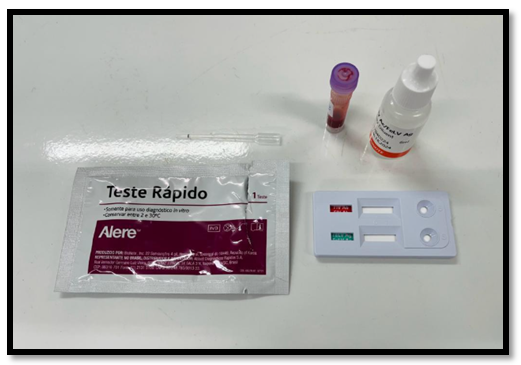
Figure 1 Alere® FIV Ac/FeLV Ag rapid immunochromatographic test (SNAP test).
Tests were conducted individually, following the manufacturer’s instructions: 10µL of blood, serum, or plasma were mixed with two drops of the dilution buffer solution provided in the kit. The mixture was then dispensed into the sample well of the SNAP device, where it migrated by capillary action through the membrane.
The results were interpreted after 10minutes. A non-reactive result was defined by the appearance of a single colored line at the control region (C), while a reactive result was indicated by two colored lines, one at the control region (C) and another at the test region (T), corresponding to the detection of antibodies against FIV and/or the FeLV antigen (Figures 2&3).
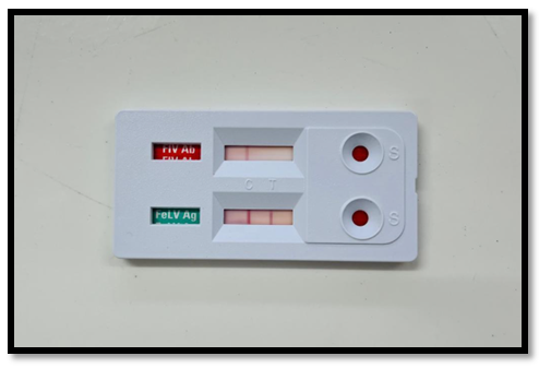
Figure 2 Non-reactive result for FIV and reactive result for FeLV.
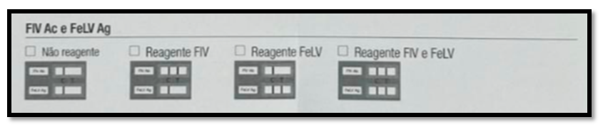
Figure 3 Interpretation guide of Alere® FIV Ac/FeLV Ag rapid immunochromatographic test (SNAP test).
This study analyzed 182 clinical records of animals that underwent rapid immunochromatographic testing for detection of viral infection. Among these 182 animals, 50 (27.4%) tested positive for at least one disease. Among the animals that tested positive, 8% (4/50) were positive for FIV only, 80% (40/50) were positive for FeLV only, and 12% (6/50) were positive for FIV–FeLV coinfection.
Cats with FeLV were assessed according to several classification criteria, as listed in Table 1: 62.5% (25/40) were males and 37.5% (15/40) were females (P=0.638). Among the animals that tested positive, 57.5% (23/40) had outdoor access (P=0.0001). When evaluated for contact with other cats, 52.5% (21/40) had regular contact with other felines (P=0.141).
|
Classification criterion |
Number infected (%) |
OR |
|
95% CI |
||||
|
Sex |
|
|||||||
|
Male |
25 (62.5%) |
1.2 |
|
0.58–2.46 |
||||
|
Female |
15 (37.5%) |
---- |
|
|||||
|
Outdoor access |
|
|||||||
|
Yes |
23 (57.5%) |
3.9 |
|
1.86–8.16 |
||||
|
No |
17 (42.5%) |
---- |
|
|||||
|
Contact with other cats |
|
|||||||
|
Yes |
21 (52.5%) |
1.7 |
|
0.83–3.47 |
||||
|
No |
19 (47.5%) |
---- |
|
|||||
Table 1 Multifactorial analysis of the main risk factors for the occurrence of feline leukemia virus in cats. Distribution of the most relevant risk factors associated with feline leukemia virus based on the classification criteria from clinical cases treated at the Veterinary Teaching Hospital (State University of the Midwest of Paraná) between January 2015 and July 2023
CI, confidence interval; OR, odds ratio.
Twelve percent (6/50) of animals tested positive for FIV–FeLV coinfection. These cases were assessed using the same classification criteria (Table 2). Notably, 83.3% (5/6) of animals with coinfection had unrestricted outdoor access, which was statistically significant (P=0.001). In terms of castration status, the majority of cats were intact (83.3%; 5/6), although this association was not statistically significant (P=0.401).
|
Classification criterion |
Number infected (%) |
OR |
95% CI |
||||
|
Outdoor access |
|||||||
|
Yes |
5 (83.3%) |
15.2 |
1.70-136.3 |
||||
|
No |
1 (16.7%) |
---- |
|||||
|
Castrated |
|||||||
|
Yes |
1 (16.7%) |
---- |
|||||
|
No |
5 (83.3%) |
2.5 |
0.28–21.8 |
||||
|
Vaccination |
|||||||
|
Complete |
1 (16.7%) |
---- |
|||||
|
None/incomplete |
5 (83.3%) |
2.5 |
0.28–21.8 |
||||
|
Contact with other cats |
|||||||
|
Yes |
5 (83.3%) |
7.3 |
0.83–64.8 |
||||
|
No |
1 (16.7%) |
---- |
|||||
Table 2 Multifactorial analysis of risk factors for feline immunodeficiency virus–feline leukemia virus coinfection in cats. Distribution of key risk factors associated with feline immunodeficiency virus and feline leukemia virus coinfection, based on clinical case classifications at the Veterinary Teaching Hospital (State University of the Midwest of Paraná) from January 2015 to July 2023
CI, confidence interval; OR, odds ratio.
Among the cats that tested positive, 83.3% (5/6) were either unvaccinated or had incomplete vaccination protocols; the vaccinations of only 16.7% (1/6) of cats were current (P=0.401). Of these animals, 83.3% (5/6) had contact with other cats, but 16.7% (1/6) were isolated (P=0.038).
The incidence of FIV in this study was only 8% (4/50), which was insufficient for meaningful statistical analysis or risk factor calculation.
The results of our study align with those of a study by Gonçalves,11 who reported an FIV incidence in male cats of 60%. The higher incidence among indoor–outdoor cats is consistent with outcomes reported by Biezus et al.,12 that 83.3% of cats had outdoor access. The outcomes on contact with other cats in the present study reflect the findings of a study by Biezus,12 namely, that 65% of FeLV-positive cats had contact with other cats.
Risk factor analysis (Table 1) revealed that male cats with outdoor access—and, subsequently, contact with stray cats—face a significantly higher risk of FeLV infection. Among all risk factors identified, outdoor access emerged as the most significant, warranting particular attention when pet owners are counseled. According to Lara et al.,13 viral circulation is significantly higher in cat populations that have outdoor access than in populations that do not. In these populations, transmission typically occurs through fighting over mates during the breeding season, territorial disputes, or food resources. Multiple studies show that the majority of FeLV and or FIV positive cats are male, with infection rates three times higher than those in females.4,5,8
Our results regarding outdoor access in FIV–FeLV coinfection cases differ from those of Cavalcante et al.,14 who reported that 60% of coinfected animals were strictly kept indoors. Our data regarding castration contrasts with that of Cavalcante et al.,14 who reported that only 31% of FIV–FeLV positive animals were uncastrated. With respect to vaccination, Vanassi et al.15 reported that 32% of respondents administered the pentavalent feline vaccine annually; however, it should be noted that this vaccine protects only against FeLV. The prevalence of contact with other cats in cases of coinfection cases aligns with that reported by Cavalcante,14 who found that 52% of affected animals lived with other cats.
Risk factor analysis for FIV–FeLV coinfection revealed that cats that had outdoor access, were uncastrated, lacked complete vaccination, and had contact with other cats faced a significantly high risk of infection for both viruses. Among these factors, outdoor access remains the most significant risk determinant. Indoor–outdoor lifestyle patterns increase viral susceptibility due to uncontrolled contact with cats that have an unknown health status.6,12,16
Mariga et al.17 at the Federal University of Santa Maria’s Veterinary Teaching Hospital reported comparable findings concerning FIV incidence, with an FIV incidence of only 4.3% (14/325) observed. Despite the high sensitivity and specificity of the Alere® FIV/FeLV Snap test (Abbott Laboratories, Chicago, Illinois, EUA) (96% and 98%, respectively), false negative test results remain possible, particularly in immunosuppressed cats or during acute infection when antibody levels are insufficient for detection.
In our case series, FeLV infection was more prevalent than FIV infection alone or FIV–FeLV coinfection. Analysis of cases with positivity revealed key risk factors, including being uncastrated, having outdoor access, and lacking complete vaccination. All these factors were significantly associated with increased susceptibility to both FIV and FeLV. These findings highlight the critical importance of owner education, particularly regarding the restriction of outdoor access, which remains the most effective preventive strategy. Given that both FIV and FeLV are associated with severe and potentially fatal health complications, prevention through responsible pet management is essential.
Not applicable.
Authors declare no conflict of interest.

©2025 Santos, et al. This is an open access article distributed under the terms of the, which permits unrestricted use, distribution, and build upon your work non-commercially.