Journal of
eISSN: 2373-437X


Case Report Volume 5 Issue 3
1Centre of Agricultural Sciences, Federal University of Alagoas, Brazil
2Institute of Biomedical Sciences, University of Porto, Portugal
3Centre Interdisciplinary for Marine and Environmental Research (CIIMAR/UP), University of Porto, Portugal
4Department of Sciences, University Institute of Heath North, Portugal
Correspondence: Themis Silva, Centre of Agricultural Sciences, Federal University of Alagoas, Brazil
Received: May 24, 2017 | Published: July 17, 2017
Citation: Silva T, Soares E, Rocha S, Santos E, Oliveira E, et al. (2017) Meglitschia spp. (Myxozoa) Infecting the Gallbladder of Eugerres Brasilianus (Teleostei: Gerreidae) from the Atlantic Coast of Maceió (Brazil). J Microbiol Exp 5(3): 00149. DOI: 10.15406/jmen.2017.05.00149
Microscopic observations using light and transmission electron microscopes are used to describe a myxosporean belonging to the genus Meglitschia Kovaleva, 1988 (Phylum Myxozoa Grassé, 1970 found infecting the gallbladder of the marine teleost fish Eugerres brasilianus (Gerreidae) collected on the NE Atlantic coast of Brazil. Typical ellipsoidal furcate and arcuate ∩–shaped myxospores, identified as belonging to the genus Meglitschia were found infecting the gallbladder and described using light and transmission electron microscopy. Mature myxospores are composed of two symmetric equal–sized valves, with two equal–sized bifurcated caudal processes (tails). The myxospores observed free in the bile measures 24.6±0.8μm long, 8.4±0.5μm wide and 5.1±0.3μm thick and are composed of two symmetric equal–sized valves, up to ~65 nm thick. Each valve possesses one opposed tapering appendage, 19.6±0.7 µm long, oriented parallel towards the basal tip of the appendages and joined along a right suture line forming a thick strand. Two spherical and equal–sized polar capsules are located side by side in the same level in the central region of the myxospores body. Each polar capsule contains a polar filament coiled in 4–5 turns. Sporoplasm is binucleated and contains the nucleus just near the polar capsules surrounded by several globular sporoplasmosomes. The sporoplasm occupies the internal portion basal of the two caudal processes.
Keywords: myxozoa, marine, fish infecting, gallbladder, ultrastructure
PFc: Polar Filament Coils; GB: Gallbladder; DIC: Differential Interference Contrast; TEM: Transmission Electron Microscopy; PC: Polar Capsules
The Myxozoa Grassé, 1970 infecting fish is an important pathogenic group having a worldwide distribution. Among these parasites the monotypic genus Meglitschia Kovaleva, 1988 has been reported infecting the gallbladder (Gb) of a marine fish from the New Zealand fauna.1,2 and a freshwater fish from Brazil, collected in a tributary of Amazon River.3 The teleostean host (Eugerres brasilianus) a commercially important fish of the family Gerreidae, common in the NE Atlantic coast of Brazil, was infected Gallbladder by myxospores of the genus Meglitschia. This genus is a small taxonomic group within the class Myxosporea that contains only two named species, M. insolita.1 (formerly described as Ceratomyxa sp.).2 from New Zealand and M. mylei.3 from Brazil. These two species were described on the base of a schematic drawing.1 and based in light and transmission electron microscopies.3 The present study provides light and ultrastructural for the description of Meglitschia sp., a myxosporean parasite infecting the Gallbladder of a marine fish on the Brazilian northern Atlantic coast. This genus, despite being similar to Ceratomyxa spp. with which it shares some morphologic aspects, differs in the typically arcuate ∩–shaped of their myxospores.
Forty specimens (27 females and 13 males) of the marine fish Gallbladder of the teleostean fish Eugerres brasilianus (Teleostei: Gerreidae) (Brazilian common name “Carapeba” or “Mojarra”) were collected during October and November 2015, from the Atlantic coast (09° 29´S/ 35° 34´W) near the city of Maceió (State of Alagoas), Brazil. Specimens measured 30–40 cm in length and weighed 600–700 g. and the respective sex register. A parasitological survey was conducted on several organs and tissues. Parasitized samples from the infected host specimens were examined and photographed using the light microscope LEICA Leitz DMBRE, equipped with differential interference contrast (DIC) optics and the digital camera LEICA DFC480 (LEICA Microsystems, Wetzlar, Germany). Measurements were taken using the software LAS V4.3 (LEICA Microsystems).
For transmission electron microscopy (TEM) were used living and refrigerated samples to obtained myxospores morphologically identified as belonging to the genus Meglitschia and fixed in 5% glutaraldehyde buffered in 0.2M sodium cacodylate (pH 7.4) for 20–24h, washed overnight in the same buffer, and postfixed in 2% OsO4 also buffered with 0.2 M sodium cacodylate (pH 7.4) for 3–4h. All these steps were performed at 4°C. The samples were then dehydrated in an ascending graded series of ethanol, followed by embedding using a series of oxide propylene and Epon mixtures, ending in EPON. Semithin sections were stained with methylene blue Azur II. Ultrathin sections were double–contrasted with uranyl acetate and lead citrate, and then examined and photographed using a JEOL 100CXII TEM (JEOL Optical, Tokyo, Japan), operating at 60 kV. No ultrastructural differences were found on myxospores obtained from the fixation of living samples and myxospores obtained from samples death some hours before (5–10h) and conserved in glace before the chemical fixation with glutaraldehyde. The observation in aquarium of the living fish does not present altered behavior. The typical morphology of the myxospores suggests that the present microparasite belongs to the genus Meglitschia, according the previous descriptions.1–4
Gallbladder and bile
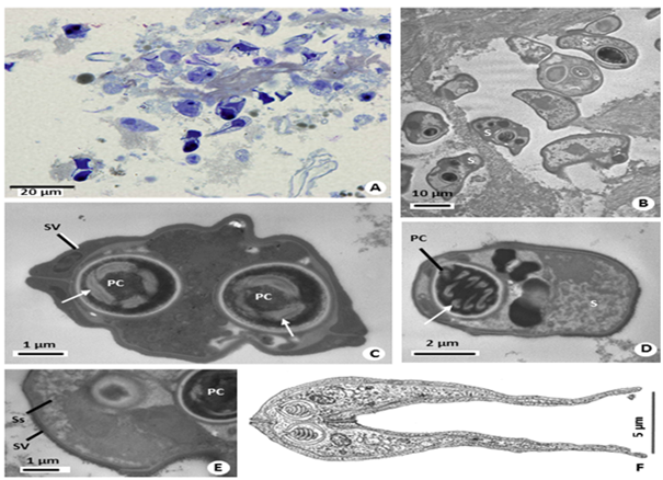
The infected Gallbladder appeared sometimes swelled and having a light brownish color when contains numerous myxospores floating in the bile. Microscopic observations revealing myxospores parasitizing the Gallbladder of some specimens (~17% infected). Numerous myxospores floating in the light brownish bile and, among them, several plasmodia containing different developmental stages were observed (Figure 1). No other organ seemed infected with same type of myxospores or plasmodia.

Figure 1 A. Semithin section showing several myxospores in the bile. B. TEM micrograph showing some Myxospores(S) sectioned at different levels. C. Transverse section showing the two polar capsules (PC) located side by side, the polar filament (arrows), and the valves comprising the wall (SW). D. Longitudinal ultrathin section of a polar capsule (PC) displaying the organization of the polar filament (arrow), and the sporoplasm cell (SC). E. Ultrathin section of the periphery of a myxospore showing the valves (SV), sporoplasmosomes (Ss), and part of the polar capsule (PC). F. Schematic drawing of a myxospore depicting its ultrastructural aspects.
Mature myxospores
The bile was collected and the isolated myxospores, as well small fragments of the Gb wall prepared for LM and TEM. Observed in LM and TEM, myxospores strongly furcate and arcuate ∩–shaped, averaging ~24µm in length, 8–9µm in width and ~5µm in thickness. Wall composed of two equal–sized valves (Figures 1A–1E), each possessing one opposed tapering appendage, ~20µm long, oriented parallel towards the basal tip of the appendages and joined along a straight suture line forming a thick strand. The strand goes around the central part of the myxospore, which in turn surrounds two equal and symmetric spherical polar capsules (PC), 2–3µm in diameter, located at the same level. Each PC contained a polar filament forming 4–5 coils. The binucleate sporoplasm was irregular in shape, contained several sporoplasmosomes and fully occupied the space of the two caudal appendages (Figures 1C.–1E). The nuclei (~1.9µm in diameter) were located near the PC. A schematic drawing of the myxospore organization, obtained from the serial longitudinal ultrathin sections, is shown in the Figure 1F.
The morphological and ultrastructural aspects of the myxospores showing a furcate and arcuate ∩–shaped organization show that these myxospores are similar with those previously described belonging to the genus Meglitschia.1–4 Based on the arc shape of the myxospore with two tapering caudal appendages oriented to the basis of spores, on the number and position of the PC and of the polar filament coils (PFc) and arrangements, the morphology of the myxospores suggested that this parasite belongs to genus Meglitschia. This myxoparasite was the first of the reported genus infecting E. brasilianus, where there is only one species of this genus (Meglitschia mylei) described parasitizing fish in Brazil.3 Based on the some morphological differences in terms of size, shape and the ultrastructural details of the myxospores and PC and PFc, when compared with those myxospores of the two species previously described, we observe that the present described isolated shows several morphologic differences to the two published species. However, the lack of the phylogenetic data on these two species does not permit the comparison with our phylogenetic results.
Upon comparison to other know Meglitschia sp., the morphometric and ultrastructural data obtained for the parasite described here, suggest that it is, possibly, to be a new member of this genus. Nonetheless, species identification is dependent of the acquisition of further information, namely at molecular level.
This work was partially supported by Engº. A. Almeida Foundation (Porto, Portugal) and Project SSPP #0067 of King Saud University. The work was partially supported by FAPEAL– Foundation of Research of Alagoas State, Brazil.
None.

©2017 Silva, et al. This is an open access article distributed under the terms of the, which permits unrestricted use, distribution, and build upon your work non-commercially.