Journal of
eISSN: 2373-437X


Research Article Volume 10 Issue 1
1 Faculty of Public Health, Universitas Muhammadiyah Semarang, Indonesia
2Health Office of Central Java Province, Indonesia
3Public Health Center of Semarang, Central Java Province, Indonesia
Correspondence: Didik Sumanto, Faculty of Public Health, Universitas Muhammadiyah Semarang, Indonesia, Tel +62 822215866 17, Fax +6224 7674029 1
Received: February 01, 2022 | Published: February 14, 2022
Citation: Sumanto D, Sayono, Meikawati W, et al. High case enterobiasis in school children and potential eggs distribution on the bed. J Microbiol Exp. 2022;10(1):33-36. DOI: 10.15406/jmen.2022.10.00349
Background: Enterobiasis caused by Enterobius vermicularis is a health manifestation of severe concern that needs to be monitored in children because of its recurrence. The cases are relatively frequent in children. The patient will experience discomfort which starts as itching in the perianal area. A decreased quality of life can also occur in children who lose nutrients due to this worm infection.
Methods: A cross-sectional study was conducted on 63 elementary school students on the outskirts of a city in Central Java province, Indonesia. Microscopic examination of perianal smear specimens and children's bedding was conducted.
Results: The incidence of enterobiasis reached 96.8% of all students under study, while the distribution of worm eggs on children's bedding reached 93.7%. The presence of worm eggs in both was significantly related (p=0.000). The incidence of enterobiasis contributed to the distribution of eggs on the sleeping bed by 49.18% (R2=0.4918)
Conclusion: Enterobiasis is a promising threat to children, and needs to be monitored through have regular annual checkups before taking treatment.
Keywords: enterobiasis, Enterobius vermicularis, periplaswab, perianal-swab, bed-swab
Enterobiasis is caused by the intestinal nematode Enterobius vermicularis (E. vermicularis).1 This type of worm is often reported to infect children,2,3 but its prevalence in adults cannot be denied.4 The incidence is quite variable in various regions, with the highest reported cases in Palestine at 98.9%.5
Enterobiasis is regarded as an issue of severe medical prominence because of its nature of instigating other diseases related to this worm infection. Case reports of appendicitis from various sites are associated with the presence of E. vermicularis.6–9 Deaths attributed to enterobiasis included a woman whose autopsies disclosed the presence of E. vermicularis localized in the duodenum and proximal ileum with intestinal bleeding.10 Ectopic enterobiasis cases have also been reported in female sexual organs such as the vagina11,12 up to the fallopian tubes13 and ovaries.14 In addition, scientific studies claim the presence of larvae in student urine samples15 which enhances the scope of re-infection after female worms lay eggs in the patient's perianal.
The incidence of infection is often associated with personal hygiene16 and bedroom sanitation conditions.17 Personal hygiene that has the most potential to be a risk factor for infection is hand hygiene and its activities related to the mouth, including fingernail hygiene,18 finger sucking habits,19 the habit of washing hands with soap both before eating and after defecation activities.16
The Indonesian government has attempted to undertake appropriate measures, including the mass worm treatment program for school children every six months.20,21 The treatment aims to kill worms in the child's body, but it should be followed by efforts to maintain the sanitation of the bedroom environment, children's playground, and healthy living behavior for children. This study aims to see the magnitude of the incidence of enterobiasis while at the same time detecting the distribution of worm eggs in children's beds and linking the two.
Study design and site
A cross-sectional study was conducted on the research subjects of elementary school children in a suburb of a city in the province of Central Java, Indonesia. All 63 students were used as research subjects.
Materials
The research materials were perianal swabs and children's bedding swabs taken with Periplaswab.22 Perianal swabs were taken in the morning after the child woke up before going to the bathroom. The child is positioned on his stomach with his pants open. Periplaswab is attached to the perianal to collect worm eggs, then fixation of periplaswab by gluing both sides of the adhesive. A bed swab was taken using a periplaswab by attaching the adhesive to several points where the child's sleeping position was on the bed sheet. Collected specimens were fixed in the periplaswab then coded and stored in a container to be brought to the laboratory before three hours.
Laboratory testing
The specimen in the Periplaswab is mounted on the reading holder and then mounted on the microscope table. Observation of the presence of E. vermicularis eggs was carried out microscopically with a fortress technique throughout the Periplaswab reading area using a Nikon Eclipse E-100 microscope with weak and medium magnification.
Ethics approval
Ethical acceptance was taken from the Health Research Ethics Commission of the Faculty of Public Health, Universitas Muhammadiyah Semarang.
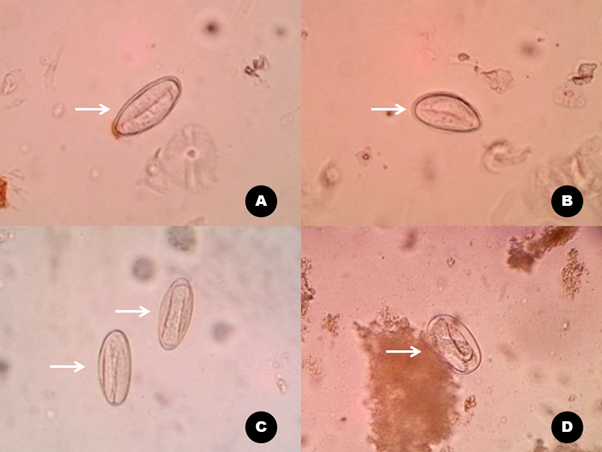
The morphology of the eggs was found according to microscopic characteristics with a typical asymmetrical oval shape. The egg wall is composed of thin transparent hyaline material that looks double. All the eggs contained embryos to larvae in a uniquely beautiful shape (Figure 1).

Figure 1 E. vermicularis egg in the typical asymmetric oval shape contains an embryo with a clear double-wall (A-B). In another position, the egg does not appear horizontal (C-D) with the embryonic development stage (C) and contains larvae ready to hatch (D).
Eggs of E. vermicularis with an average of 45 eggs per small visual field were dense and filled the entire visual field of microscopic observation. This view is the case with the highest degree of infection (Figure 2).
The prevelence of Enterobiasis was found to be in high number accounting to more than 90%. This result is an important finding for control efforts. Information on the presence of worm eggs scattered in infected children's beds is a good basis for parents to change bed linen regularly.The two are significantly related (Table 1).
Specimen |
Positive |
Negative |
Total |
p-value |
Perianal swab |
61 (96,8%) |
2 (3,2%) |
63 (100%) |
0,001 |
Bed swab |
59 (93,7%) |
4 (6,3%) |
63 (100%) |
Table 1 E. vermicularis eggs on perianal swab and bed swab
There was an interaction between the incidence of the children infection and the worm eggs presence in the sleeping bed. It is shown in a trend graph with R2 = 0.4918 (Figure 3).
The morphology of the eggs of E. vermicularis was found to be asymmetrical oval in shape and double transparent hyaline walls comprising of embryos to larvae with a distinctive shape.23 Microscopically, infective eggs will show larvae that are actively moving in the eggshell.24 The number of eggs observed in each field indicates the degree of infection. The degree of infection was proportional to the number of eggs per field.
The finding of a very high incidence of enterobiasis is surprising. The higher rate of its prevalence makes this disease an issue of medical concern. This finding is the second-highest case report ever reported after the case in West Bank Palestine at 98.9%.5 Similar cases have been reported in Al-Hilla city-53%,25 Grobogan Indonesia-52.6%,26 Kalar, Iraq-24.9%,27 Babol Iran-22.2%,28 Subang Indonesia-13.9%,29 Mazandaran Iran-7.1%,30 Sabzevar Iran-3.49%,31 and Bulgaria-0.91%.4
B. vermicularis can attack all age groups, but the data reported are more common in the pediatric group.17 The reports from Bulgaria stated that cases in children were significantly more common than in the adult age group.4This is related to personal hygiene behaviors such as finger sucking habits,32 not washing hands before eating16 and after defecation.33
Worm eggs that stick to fingers and fingernails due to contact with the rectal area or worm eggs on the distribution media if not cleaned have the potential to become an intermediary for the entry of worm eggs into the mouth34 which is the initial stage of infection in enterobiasis cases.35 The eggs will enter the digestive tract and hatch in the small intestine, then the larvae will become adults in the colon.1 Transmission through the air by inhalation is very likely to occur, this is related to the habit of shaking the child's sleeping blanket before folding it back in the morning.17
The discovery of worm eggs on the child's bedding indicated that the child's perianal area had undergone intervention after the female worm laid eggs. The interventions that occur can be intentional or unconscious when the child sleeps at night. The presence of discomfort in the perianal as itching will encourage efforts to scratch the perianal area using your hands. This activity, both from outside the pants and from the inside, has the potential to cause eggs to fall on the child's underwear and spread on the bed. Children who sleep without trousers are more likely to spread eggs on the bedding. Similar studies involving the bed variable reported that the sleeping bed was significantly associated with the incidence of Enterobiasis.33,36
The existence of a significant relationship between the incidence of enterobiasis in children and the presence of worm eggs on bedding is easily understood. This means that worm eggs can be spread on bedding only if the user has enterobiasis. The logic of the existence of sufferers causes the presence of worm eggs, of course, it is undeniable. The R square = 0.4918 means that the distribution of E. vermicularis eggs on the bed is supported by 49.18% by the incidence of enterobiasis in children, the rest is influenced by other factors. Other factors that need to be observed in further studies include perianal scratching behavior, children's sleeping habits, finger sucking habits, and other variables related to the sanitation of children's bedrooms.
Enterobiasis is an infectious disease that compromises children from normal well being. Finger hygiene includes nail care and the habit of washing hands with soap are important factors in preventing infection. The sanitary condition of the child's bedroom must be considered, especially the cleanliness of the bedding.
Thank you to the Laboratory of Epidemiology and Tropical Diseases, Universitas Muhammadiyah Semarang for facilitating laboratory testing.
Authors declare that there is no conflict of interest.

©2022 Sumanto, et al. This is an open access article distributed under the terms of the, which permits unrestricted use, distribution, and build upon your work non-commercially.