Journal of
eISSN: 2373-437X


Review Article Volume 1 Issue 3
School of Chemical and Biotechnology, SASTRA University, India
Correspondence: Ranganathan Vasudevan, School of Chemical and Biotechnology, SASTRA University, Thanjavur - 613 401, India, Tel 91-8121119692
Received: June 07, 2014 | Published: June 18, 2014
Citation: Vasudevan R. Biofilms: microbial cities of scientific significance. J Microbiol Exp. 2014;1(3):84-98. DOI: 10.15406/jmen.2014.01.00014
Biofilms are defined as the self produced extra polymeric matrices that comprises of sessile microbial community where the cells are characterized by their attachment to either biotic or abiotic surfaces. These extra cellular slime natured cover encloses the microbial cells and protects from various external factors. The components of biofilms are very vital as they contribute towards the structural and functional aspects of the biofilms. Microbial biofilms comprises of major classes of macromolecules like nucleic acids, polysaccharides, proteins, enzymes, lipids, humic substances as well as ions. The presence of these components indeed makes them resilient and enables them to survive hostile conditions. Different kinds of forces like the hydrogen bonds and electrostatic force of attraction are responsible for holding the microbial cells together in a biofilm and the interstitial voids and the water channels play a significant role in the circulation of nutrients to every cell in the biofilm. The current review adds a note on bacterial biofilms and attempts to provide an insight on the aspects ranging from their harmful effects on the human community to their useful application. The review also discusses the possible therapeutic strategies to overcome the detrimental effects of biofilms.
Keywords: bacterial biofilms, applications and antibiotic resistance, formation and development, therapeutic approaches
MIC, microbial influenced corrosion; MBC, minimum bactericidal concentration; MBEC, minimum biofilm eradication concentration; Fg, fibrinogen; Fn, fibronectin; Cn, collagen; MSCRAMMs, microbial surface components recognizing adhesive matrix molecules; PIA, polysaccharide intercellular adhesin; CWA, cell wall-anchored; Aap, accumulation-associated protein; Bap, biofilm-associated protein; Bhp, biofilm-associated homolog protein; Embp, extracellular matrix binding protein; PSMs, phenol-soluble modulins; NIH, national institute of sciences.
Centuries have passed since the discovery of microorganisms and various attempts have been made to substantiate the outcome of many research investigations in favor of science and technology. Since the time of the discovery of bacteria, the progressing era has witnessed a wide range of developments in terms of experimental investigations and research analysis which involves proper planning, implementation and applications that can be fruitful to the mankind. Nevertheless, life science is an enormous area that comprises of various allied and distinct fields which in turn makes the scientific investigators enthusiastic to see the sights and the times gone by has indeed seen many such pioneers. However, it wouldn’t be possible to exemplify all the associated facets of life science, the current review attempts to illustrate the concept of biofilms which are considered as a vital part associated with the microorganisms and endeavors to disclose the importance of this biological component and their application in research. It is a well known fact that the beginning of the seventh century has provided the world with an insight of microorganisms and the credit goes to Antonie Van Leeuwenhoek who first observed these creatures in the calculus of his own teeth.1 These deposits comprised of different forms of ‘animalcules’ which are known to the current world as bacteria of dental plaque. Indeed the presence of such dental plaques is regarded as the earliest evidences to validate the existence of bacterial biofilms. In the present day, biofilms are defined as microbial communities attached to a surface that can either be biotic or abiotic. In addition, biofilms can also be found in submerged and humidified conditions. Their presence cannot be confined to solid substrates but they can be found as a floating mat on liquid surfaces.1,2 Biofilms can also be found on a variety of other surfaces like natural aquatic systems living tissues, medical devices like indwelling catheters and industrial piping systems. They are self produced extra cellular slime comprising of microbial communities and are primarily composed of water and polysaccharides, besides the presence of other vital macromolecules like nucleic acids, proteins, enzymes and lipids.
Biofilm research also encompasses the concept of biofilm engineering which illustrates the biofilm bases technologies and their applications. This research has in turn enabled the researchers and scientific investigators to study the influence of biofilm deposits on the metals in order to quantify the mechanism of microbial influenced corrosion (MIC). Many researchers have been successful in determining the minimum concentration of any drug that can be effective in controlling or inhibiting or eradicating the biofilms which is referred to as minimum inhibitory concentration (MIC), minimum bactericidal concentration (MBC) and minimum biofilm eradication concentration (MBEC).3 In addition, the employment of computerized techniques to validate the significance of biofilms cannot be contradicted. Indeed, the use of such sophisticated techniques has revealed several interesting facts about biofilms and has provided the scientists and research investigators with an in depth view on the biofilms which enabled them to characterize the different complicated aspects which could not be possible through conventional methods. However, biofilm research primarily involves three significant stages which can be listed as the identification and characterization of the biofilms, followed by their applications which are considered to be multidirectional. It is a widely accepted fact that biofilms serve several purposes of microorganisms that are embedded in it and confer them with specialized functions that makes them pathogenic and resilient to the commonly employed drugs. In addition, biofilms are considered to have various applications that are beneficial to industries and environment which makes it a core topic of research.
Biofilms are considered as vital structures associated with microorganisms composed of a variety of bio molecules and are usually defined as extra polymeric substances or matrices which comprise of a group of microorganisms and are known to confer the microorganisms with specialized functions which are not processed by the non biofilm forming microorganisms or the free floating planktonic microorganisms.4 They are self assembled microbial structures that are capable of optimizing their functions and are known to regulate various metabolic activities in favor of the enclosed microbial communities. Organisms embedded within the biofilm matrix are not scattered but are arranged systematically and are regulated through a series of genes that result in the formation, multiplication and dispersal of the mature biofilms. These biofilms are the self produced hydrated extra polymeric matrix which forms the immediate environment of the microorganisms.4 In fact, many scientific investigators have regarded biofilms as an integral component of a microorganisms rather than considering it as an external component because it promotes many regulatory and metabolic activities among the microorganisms and it provides the organism with the required nutrients for their survival and enables the organism to overcome adverse environmental condition as a consequence of nutrients scarcity. Numerous studies in the past have attempted to understand the biofilms and their significance until 1978 when scientific studies revealed the existence of bacteria as sessile communities within an enclosed matrix.5 Earlier studies on dental plaques has allowed the researchers to conceive the mechanism by which the microorganisms attach to solid or liquid surfaces and derive the benefits from the surroundings for its survival.
Scientific investigations have characterized biofilms as the sessile communities enclosed within a bio polymeric inclusion attached to a living or a non living substance. The microbial communities within the enclosure comprises of an altered phenotype which in turn physiologically demarcates them from planktonic microorganisms.6 Clinical studies have confirmed the role of biofilms in causing human infections which accounts up to 60%.7 Biofilm formation and development is a complex mechanism and a dynamic process which provides a better understanding on biofilms and will lead to novel therapeutic approaches. Despite the fact that, research studies validate the significance of biofilms in various allied areas of life science, the darker aspect cannot be contradicted. Biofilms are associated with numerous chronic infections that are capable of claiming a patient’s life and their role in infecting the biological devices among hospitalized patients is a universally accepted fact. Biofilms are also found on various biomaterials used in medicine such as urinary catheters and orthopedic devices.8 Another major aspect that concerns the global scientific community is the extent of virulence exhibited by the microbial population embedded within the polymeric matrix and demonstrative studies have validated the coordination between biofilm formation and quorum sensing mechanism among pathogenic bacteria.9 In addition, the other significant aspect that has challenged the scientific researchers worldwide is the extent of antimicrobial resistance shown by the pathogens within the extra polymeric matrix which makes the pathogen resilient to the commonly employed drugs.10,11 Nevertheless, the genetics as well as the environment are important aspects that determine the nature of biofilms. In fact, the environmental condition is one significant factor that makes the pathogen flexible towards a variety of surroundings and genetic diversity is equally vital and this diversity has in turn resulted in the development of new strains as a consequence of horizontal gene transfer.12,13 Biofilms are regulated by a variety of genetic and environmental factors and is in fact the major means of infection among human beings.
What are biofilms composed of?
It is a widely accepted fact that biofilms comprises of bacterial cells and extra polymeric substances but the vital ingredients that are present with in a biofilm matrix have drawn the attention of many scientific leaders all over the world. These extra polymeric enclosures not only favor the microorganisms by providing the essential nutrients but also create a favorable environmental condition for their survival and offers architectural integrity.14 In addition, it enables genetic transfer and intracellular communication. Therefore the composition of biofilms is very significant as they promote different metabolic and physiologic activities at various levels. Research studies have regarded biofilms as organized systems with suitable conditions which provides the bacterial with structural and functional merits.15,16 Structural studies on biofilms have revealed the presence of microbial cells and extra polymeric substances that accounts to 50 – 90% of their total organic carbon and can be regarded as the major component. Despite the fact, the extra polymeric substances differ in their physical and chemical properties; they are mainly composed of polysaccharides. In Gram negative bacteria, the biofilm polysaccharides can either be polyanionic or neutral. The presence of uronic acids like glucuronic acid, galacturonic acid and mannuronic acid offers the characteristic anionic property to the extra polymeric substances.17 Divalent cations like calcium and magnesium maintains the structural integrity by holding the polymers together and provides the binding strength for the biofilm development. However, bacteria like Staphylococcus are known to have cationic chemical composition and demonstrative studies on coagulase negative Staphylococcus have shown the presence of teichoic acid in combination with small amounts of proteins.17,18 The extra polymeric matrices that enclose the microbial community are highly hydrated due to large amounts of water and this in turn favors the hydrogen bonding between the embedded microbial cells. The primary configuration of the bacterial biofilms is indeed determined by the composition and structure of the polysaccharides. Many bacterial extra polymeric substances comprises of hexose residues as their backbone which tends to make them rigid and in turn results in poor solubility.19 The amount of extra polymeric substances significantly varies among different organisms and the quantity increases as the age progresses.20 In addition to polysaccharides and metal ions, the bacterial biofilms comprises of bio molecules like DNA, protein, lipids and organic substances (Figure 1). Excessive amount of carbon and reduces rates of nitrogen, potassium and phosphates inhibits the production of biofilms and in contrast, slow bacterial growth enhances the formation of biofilms. In addition, presence of hydrated environment avoids the harmful effects of desiccation in natural conditions. Biofilms consists of micro colonies of bacterial cells that are separated by water channels.21 These water channels allow the flow of nutrients, oxygen and microorganisms from one site to other through fluid circulation and they also maintain the hydrated condition which provides a natural environment for the survival of the enclosed microbial community. Biofilms are highly intricate and are usually heterogeneous in nature which comprises of thin base deposits ranging from monolayer to several layers of cells consisting of water channels.14 The type of organism also influences the structure of biofilms. An important aspect that influences the microbial community that is enclosed with in a biofilm is its thickness. Research studies and scientific investigations have shown that the pure cultures of bacterial species like Klebsiella pneumoniae and Pseudomonas aeruginosa exhibited a thickness of 15 and 30μm where as a mixed culture of these species displayed the existence of thicker biofilms when compared to their pure cultures. Research investigations have also substantiated the beneficial effects of mixed culture biofilms where one species enhances the stability of the other.22,23
Based on the studies performed in the past and the ongoing research it is understood that the biofilm structural design is heterogeneous and relies on the kind of bacterial community embedded within it due to factors like genetic diversity which vary from one organism to another. In addition to genetic diversity, the environmental conditions are equally vital and signify the nature of biofilms. The Table 124 displays the various components of a biofilm with their respective percentages.
S. No |
Component |
Percentage of matrix |
1 |
Water |
Up to 97% |
2 |
Microbial cells |
2-5% |
3 |
Polysaccharides |
1-2% |
4 |
Proteins |
<1-2% (includes enzymes) |
5 |
DNA and RNA |
<1-2% |
6 |
Ions |
Bound and free |
Table 1 Composition of biofilm24
It is evident from the Table 124 that the biofilms not only comprises of microbial cells and polymeric matrices but in addition it consists of a variety of bio molecules including proteins, enzymes and ions. The major component of the biofilm is water which in turn enables the easy flow as well as access of the nutrients for the enclosed microorganisms. Indeed, these different components signify the integrity of biofilms and make them resilient towards a variety of environmental factor. When environmental factors are considered, these microbial biofilms can be found on inundated conditions and submerged platforms in natural and industrial environment.25 Despite the fact, that the biofilms are mainly composed of extra polymeric matrices, the importance of DNA cannot be denied as it is essential for the establishment of the biofilm structure.26 Therefore it is understood that the each single component of a biofilm contributes towards the survival of the embedded microbial communities and in turn regulate a variety of metabolic activities.
The extra polymeric substances offer protection from a range of antimicrobial agents and the bio molecules like proteins, lipids and nucleic acids enhances the mechanical stability of the biofilms and allows the microbial communities to attach to the a variety of surfaces ranging from biotic to abiotic in nature including medical devices like catheters and other artificial valves. Research studies have confirmed that the biofilms are highly complicated and form a cohesive three dimensional structure that is organized and interconnected with specialized functions.4
Biofilm formation in gram positive bacteria: Biofilm formation and dispersal is a complicated process which involves a series of vital factors and the mechanism of dispersal occurs after the maturation of the biofilms. In fact, biofilms have been regarded as the main source of infection and their development involves different stages like primary attachment, accumulation, maturation and dispersal.27 Despite the fact, that the stages involved in the process of biofilm formation are same, this section of the review discusses the stages of biofilm formation in Staphylococcus species. The various stages involved in the process of biofilm development and dispersal are as follows:
Gram negative bacteria biofilm formation: The previous section has emphasized on the various stages of biofilm formation in Gram positive bacteria and the current section will add a note on the Gram negative biofilm formation. It is a widely accepted fact that Escherichia coli is known for its capability of forming biofilms and has been targeted by many researchers to understand and reveal the mechanism. It is a facultative anaerobic bacterium of the gastrointestinal tract and employs a variety of extracellular appendages for its colonization and development.46 The stages involved in the biofilm formation are as follows.
Curli otherwise referred as culi fimbria is a thin aggregative external appendage that is a feature of the members of the Enterobacteriaceae which includes pathogens like Shigella, Citrobacter and Enterobacter. They promote the cell adhesion by attaching to the proteins of the extracellular matrix such as fibronectin, laminin, and plasminogen. Curli fimbria also enhances the attachment to the abiotic surfaces by promoting cell to cell interaction.64 The csgBA operon and csgDEFG operon comprises of the genes involved in the production of curli fimbria. The production of curli fimbria is highly regulated and occurs at temperatures ranging from 28°C to 37°C depending of the kind of isolate. However, the expression of curli is certain strains of the pathogen remains mysterious and is yet to be unraveled.
Conjugative pili are hair like external appendage which enables the transfer of DNA through the process conjugation and their role in enhancing the biofilm formation has been validated by prior demonstrative studies. Mixed cultures of E. coli K-12 strains (poor biofilm producers) and E. coli communities of conjugative plasmid have enhanced the biofilm forming capacity of the K-12 strains of E. coli.65 The initial attachment and biofilm colonization in a nonspecific manner on abiotic surfaces is promoted by the F pilus which enables the cell to cell contact and stabilizes the biofilm structure. Studies have also confirmed the importance of conjugative and non conjugative plasmid in promoting biofilm formation due to the presence of factors favoring biofilm development.66–68 Several cell adhesion proteins encoded by genes have been identified in pathogenic E. coli that favors biofilm formation.69
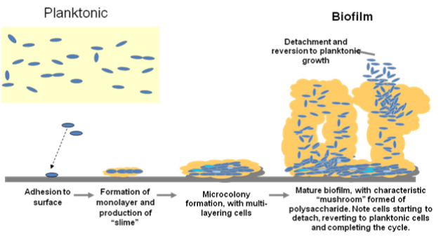
Once the desired population of the cell is attained, the mature microbial cells are detached and dispersed in to the environment. This mechanism is cyclic as the released microbial cells from a mature biofilms attach to new surfaces and the same process continues. The following diagram depicts the various stages of biofilm formation.
The Figure 271 is a diagrammatic representation of the formation of biofilms which begins from the preliminary attachment of the planktonic forms to a surface followed by their adhesion to the surface. At this stage the attachment is reversible. Further stages results in the formation of monolayer of cells and the production of the extra cellular slime begin. The proceeding phase witnesses the formation of microbial communities with multi layer of cells enclosed within the extra polymeric matrix and at this stage the attachment becomes irreversible. The further development of biofilms results in their maturation followed by the process of detachment and dispersal. It is a widely accepted fact that a biofilm comprises of microbial cells and its major constituent is water which in turn favors the attractive forces like hydrogen bonds which exist in between the cells embedded in the self produced slime and maintains a hydrated environment to favor the growth and development of the microbial cells within the biofilm. Presence of water channels plays a crucial role in the distribution of vital nutrients required for the growth of the microbial cells. In addition to the polysaccharide which is the main component of the extra cellular matrix, the biofilm also comprises of macromolecules such as DNA, RNA, lipids, proteins, enzymes and ions which equally contribute towards the metabolic processes that are essential for the existence of the microbial biofilm and macromolecules also play a significant role in maintaining the stability of the biofilm structure. In fact, microbial biofilms have lead to the development of new strains as a consequence of horizontal gene transfer and the inclusion of a variety of genetic and environmental factors has made the bacterial biofilms highly complex.

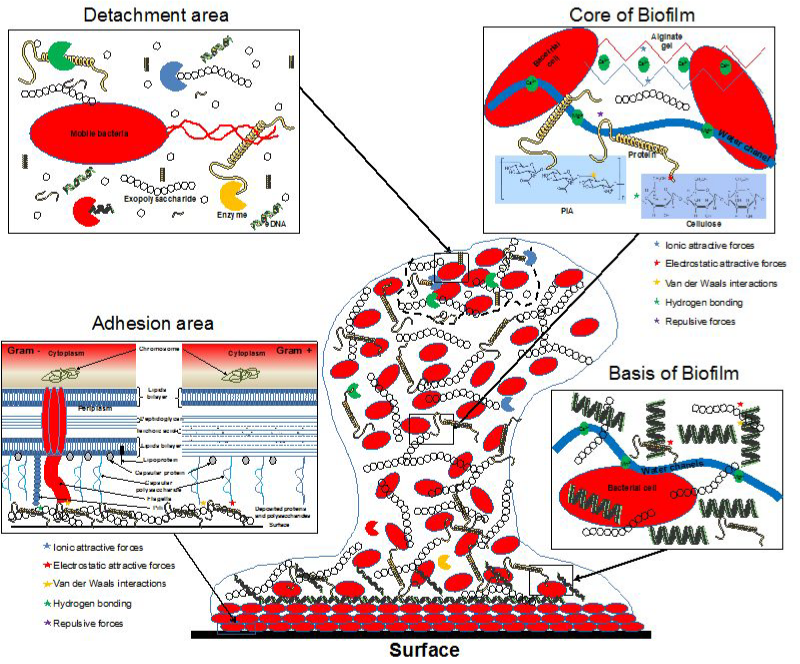
The Figure 327 tries to provide an overview of a mature biofilm and different kinds of bonds responsible for the structure and stability of the biofilm. The central part of the figure represents a mature biofilm accumulated with microbial community composed of multilayer of cells and is on the verge of dispersal. The bacterial cells are attached to a solid substrate and are enclosed within the extracellular slime. The contact of the bacterial cells with the solid substrate is promoted by various external appendages like fimbria and pili. Genetic factors are equally responsible as the controlled expression of certain set of genes decides the nature of biofilms. The extracellular components comprises of DNA in addition to polysaccharides and proteins. The interior part of the biofilms comprises of the water channels that enable the supply of ions and nutrients to the microbial community. The detachment of the microbial cells is a consequence of certain microbial enzymes that tear down the extra polymeric matrix resulting in the dispersal of the microbial cells and enables the microbes to colonize new surfaces.

The previous sections provided an overview of biofilms, their structure and the different stages involved in the formation and development of bacterial biofilms. It emphasized on the microbial communities of Gram positive and negative bacteria enclosed within the extra polymeric slime and highlighted the importance of various components in maintain the structure and integrity of the biofilms. The current section endeavors to add a note the importance of biofilms and their applications. Major microorganisms of natural, industrial and clinical origin are initially confined to a surface for their growth and development and are released in the environment after their growth and maturity. Therefore studies have defined biofilms as structural and functional microbial communities with specialized characters found on natural and artificial surfaces.72 Advancements in the allied and distinct areas of Science and technology have enabled the pioneers of various fields ranging from Biotechnology to medicine to carry out an interdisciplinary research to explore the significance of bacterial biofilms. Applications of biofilms can be of industrial and ecological significance and range from the treatment of industrial and waste waters to the decontamination of the polluted sites. Studies have also shown the ability of bacterial biofilms in degrading the industrial contaminants of chemical origin which are considered to be recalcitrant by using them as carbon source.73 Many researchers have put forth their research outcomes and it was evident that the extent of biofilm formation relied on the interaction between the microbial community and the specific surface. The eminent scientists and researchers feel that the wild strains are competent in terms to biofilm formation when compared to laboratory strains. It is believed that the bacteria are known to adhere to a variety of surfaces due to their metabolic activities and this phenomenon is prominent among the wild types which in turn signify the importance of wild type strains.74 Research studies have confirmed the role of biofilms as an important component of food chain in water bodies like rivers and stream as they serve the feeding purpose of invertebrates which in turn are consumed by the fishes of the aquatic system.75
Biofilms can be of industrial importance and can be used for constructive purpose of industrial appliance. Sewage purification process is an example that validated the significance of biofilms. The treatment of sewage water involves a phase where the contaminated water is allowed to flow over the filters consisting of layer of biofilms which confirms the constructive purpose of bioflms. When the contaminated water flows over the filters consisting of microbial biofilms, the nutrients from the flowing water are extracted by the microbial biofilms and they play a vital role in the removal of the organic matter from the contaminated water.76 The major factor that influences the treatment of water through biofilms is the surface area. In addition to its beneficial effects in treating contaminated water and polluted sites, microbial biofilms play a vital role in breaking down the unwanted debris formed from the dead fish and aquatic plants and absorb the heavy ions from the water without depleting the oxygen content and in this manner it contributed positively towards the ecological balance.77 The composition of the biofilms is equally significant as every component contributed towards the specialized functions performed by these microbial communities within biofilms. The extracellular slime extensively varies in its composition, structure and properties which in turn make it difficult to go over the main points in terms of their contribution.74 Species like Bacillus cereus, B. licheniformis, Arthrobacter species, Pseudomonas species, Candida albicans etc are known to produce several strains that are capable of forming the biofilms on a variety of substrates and are of industrial importance.78,79 Research studies have confirmed the adhesion capacity of Arthrobacter oxydans 1388 to various polymeric substrates including acrylamide and cellulose acetate.
The available scientific data on the adhesion of biofilms also signifies the importance of various factors like the nature of the cell surface, age of the cell culture, ions and the type of polymeric matrix. These factors indeed decide the extent of adhesion of the microorganism to the substrate and alterations in these factors can lead to desorption of the microbial community which in turn influences their productive aspects.80 There are several advantages that favor the bacterial communities that are enclosed within the extra polymeric matrix and the extent of competence and complexity enhances as the stages progresses. In fact, the planktonic forms that produce the biofilms are less virulent in the initial stages when compared to the mature biofilms of the same species which are highly virulent.
Lower concentrations of phenol are toxic to human society and can lead to dire consequences and demand the requirement of appropriate methods to overcome the difficulty as there is a need to reduce the phenol content from the natural environment as a consequence of industrial practices. Microorganisms can be employed as an option to counteract the problem of phenols as many studies have confirmed the role of microorganisms in degrading phenols. In fact, the biological treatment of water has become an attractive means as phenols are reduced to harmless products and secondary mineral wastes by the action of microorganisms.81 Several scientific studies in the past have attempted to validate the importance of microbial biofilms in reducing the harmful impact of phenols which involves microorganisms like Arthrobacter species, Pseudomonas species, Acinetobacter, Candida through aerobic degradation of phenols. The demonstrative studies on the aerobic degradation of phenols by microbial biofilms were time specific and different strains of pathogens showed the time specific and dependent efficacy in the degradation of phenols.82-87 Attempts have been made to explore the mechanical properties of microbial biofilms which demonstrates the rheological behavior of fluids. Investigation and understanding of these properties provide an insight on the basis of the microbial biofilms of industrial and medical importance. Research studies have confirmed the existence of viscoelastic properties among the biofilms of Pseudomonas aeruginosa and the studies attempts to signify the importance the surface tension of the solid surface. Available scientific data and research reports substantiate the presence of organic polymer properties among the biofilms of Streptococcus mutans on dental plaques.88,89
Bacterial association with humans has been dated back to centuries and studies have revealed their consequences towards the human society. However, all the microorganisms are not pathogenic but their presence in all type of environment creates an alarming situation among humans. Several demonstrative studies have revealed an existence of complex interaction between the factor of an individual’s own immune system and the bacterial pathogens forming biofilms.90 Nevertheless, the microorganisms as a community have several advantages when compared to a single microbial cell and this I turn makes them resilient to host immune factors. The pathogenic microbial communities are capable of causing chronic infection n humans that can tolerate standard treatment. Microbial communities within the extra polymeric matrix are highly protected from a variety of factors and enable the embedded pathogens to survive extreme conditions. Many demonstrative studies have confirmed the role of bacterial biofilms in compromising an individual’s immune system and is capable of resisting the factors of the host immune system.90 This is one of the reasons that infections as a consequence of biofilms are rarely resolved by an individual’s own immune system. The extra polymeric matrix is the first means of defense in favor of the pathogen and the presence of exopolysaccharide alginate protects the microbial cells from the process of phagocytosis where the macrophages and neutrophils fail to engulf the microbial cells.1,91
Biofilms and antimicrobial resistance
Biofilms have in turn enhanced the resistance among the microbial community towards a variety of antimicrobial agents in addition to their resilience to various host factors. One of the major reasons for the increase in resistance to antibiotics is the expression of different genes that encodes a set of protein that confers the microbial community with this character. In addition to genetic factors which involve the expression of vital genes in favor of the microorganisms, the extra polymeric matrix prevents the penetration of the antimicrobial agents in to biofilms and quorum sensing is another vital factor that favors the microbial communities within biofilms.92 The biofilm structure enables the pathogens to tolerate the antimicrobial agent which is an in built character of the bacterial biofilms.93 The reduced ability of the employed antibiotics to break through the microbial biofilms is considered to be a crucial reason for the increase in the antimicrobial resistance. This could be a consequence of chemical interactions that occurs within a microbial biofilm or due to the existence of anionic polysaccharides. Studies carried out on the biofilms of P. aeruginosa have revealed the significance of alginates that are capable of binding to positively charged amino glycosides and prevents their penetration in to the biofilms.94 Studies carried out on coagulase negative Staphylococci have shown the importance of extracellular matrix in offering resistance against glycopeptides antibiotics in planktonic cultures. In addition, scientific investigations and demonstrative studies have confirmed the existence of mechanism within the biofilms that are capable of sequestering the antimicrobial agents and prevent them from reaching their target site.95,96 Several pathogenic bacterial strains have developed resistance against β-lactum antibiotics like penicillin and cephamycin due to the presence of β-lactamase enzyme that gets accumulated in the bacterial biofilms due to secretion or cell lysis. However, the extra polymeric matrix cannot be considered as the sole factor that prevents the entry of the antimicrobial agents in to the biofilms due to the matter of fact that there are other intrinsic factors that cannot be contradicted.97 Research studies have highlighted the importance of slow growing bacterial cells and have confirmed their tendency to escape the activity of the antimicrobial agent because the antimicrobial agents are highly effective on actively growing cells and this property of slow growth enhance the scope of resistance among the pathogenic bacteria.98 Biofilms enhances the ability of the microbial communities to adapt adverse conditions and this is an important aspect that makes the pathogen resilient to many external factors including antimicrobial agents.99 Microbial community within the biofilms can go in to dormancy during the adverse condition which in turn enables the pathogens to persist in hostile environment. These cells are referred to as persister cells which represents the small population of microbial community that are inactive and are highly protected. These cells accounts to 0.1% to 10% of the biofilms which are capable of escaping the activity of the antimicrobial agents and they serve as the initiator cells to further resume the process when the conditions become favorable.100–102 Expression of several phenotypic and genotypic factors followed by the microbial attachment to the surface makes the biofilm forming bacteria more virulent when compared to the planktonic forms and confers them with the ability of tolerating the antimicrobial impact. These virulent biofilm phenotypes are capable of expressing periplasmic glucans that bind to the antibiotics and physically sequester them which in turn reduces the efficacy of the antibiotics.103,104
Altered expression of genes or stress response within the biofilms can reduce the antimicrobial susceptibility among the microbial community and in turn enhances the resistance. Antibiotics target the specific sites and studies have confirmed that the biofilms employs specific genes capable of altering these target sites to protect the microbial community within the biofilms.
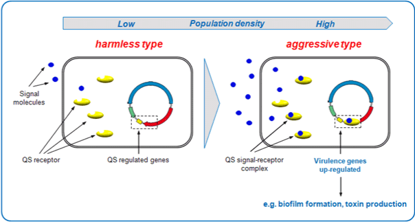
Quorum sensing and biofilms (Figure 4)105 are intimately related terms which are coordinated through a set of expressions of genes. In fact, quorum sensing process coordinates the mechanism of biofilm formation. Scientific investigations have enables the researchers to explore the significance of quorum sensing in biofilm formation. Quorum sensing can be defined as cell to cell signaling involving cell communication and it has shown to play a significant role in the formation of biofilms among pathogenic bacteria. It is a vital process which monitor the bacterial cell population and when the required cell density if attained it results in the formation of signaling molecules known as autoinducers which favors the mechanism of quorum sensing.106 Higher density of bacterial cell population enables the pathogen to secrete the autoinducers in to the external environment which in turn triggers the quorum sensing mechanism. Alteration of the quorum sensing mechanism by the process of enzymatic degradation prevents the formation of biofilms or weakens the established biofilms. Quorum sensing also plays a significant role in regulation of gene expression as well as the cell density dependent signaling mechanism. When the required cell density is achieved, the autoinducers are secreted which bind to the transcriptional factors and cause the activation or the suppression of certain genes that could be beneficial to the pathogen.107 The autoinducers known to trigger the quorum sensing mechanism significantly vary among Gram negative and positive bacteria. The Gram negative bacterium employs N-homoserinelactone which is a protein and the length of the antoinducer relies on the extent of cell density. In contrast, the Gram positive bacterium makes use of peptides as signaling molecules to initiate the quorum sensing process.
Studies have confirmed the importance of quorum sensing in increasing the scope of nutrient availability and make the bacteria highly competent against the other competing bacteria and the environment.108 Scientific investigations and demonstrative studies have validated the importance of quorum sensing in bacterial biofilm formation. Genetic experiments involving the mutant strains of bacteria have been performed to illustrate the importance of cell signaling and communication in the formation of biofilms. Mutant bacterial strains lacking the vital gene for the production of signaling molecules were used and the extent of biofilm formation was observed in order to signify the importance of quorum sensing in bacterial biofilms.109 Despite the fact, the studies have substantiated the significance of quorum sensing in bacterial biofilm formation, there are certain studies rules out the significance of quorum sensing and have indicated that quorum sensing does not influence the biofilm formation.110 However, the knowledge of signaling molecules allows the recognition of the compound that is capable of altering the quorum sensing associated processes and this in turn can have an influence on the extent of biofilm formation.111 The information on the chemical structure of the signaling molecule is very vital to understand the importance of cell signaling and will signify the role of quorum sensing in bacterial biofilm formation. The identification of suitable targets enables the development of innovative strategies that can be employed to control the harmful effects of biofilms since many studies and demonstrative experiments have intimately related the concepts of quorum sensing and biofilm formation.112 However, further studies are required to validate the significance of quorum sensing in bacterial biofilm formation and the importance of cell signaling in influencing the virulence and the extent of antimicrobial resistance exhibited by the pathogens.

The spread of a wide variety of infections as a consequence of bacterial biofilms has in fact challenged the clinical society. Studies carries out at the National Institute of Sciences (NIH) has regarded biofilms as a main consequence of infections which accounts to about 60%.113 Scientific investigations and demonstrative studies have also confirmed the role of biofilms in conferring gingival infections in adults which accounts to around 40-50%.114,115 Studies have also revealed the presence of biofilms related infections among the infants diagnosed with cerebrospinal- fluid shunts which accounts to 15-20%.115 Majority of patients hospitalized for treating urinary tract infections are liable to encounter the harmful consequences of biofilms infections and the incidence was higher among the patients with urinary catheters. Studies have shown that the extent of antibiotic resistance exhibited by the catheters associated biofilms were high and were difficult to treat.116 The indwelling catheters can be infected by Gram negative and positive bacteria which include pathogens like E. coli, P. mirabilis, P. aeruginosa, S. aureus, Enterococcus faecalis etc. The bacterial biofilms found on the indwelling urinary catheters usually comprises of a single species but as the time advances it develops in to a biofilm comprising of mixed species of pathogens. The biofilm infection among the patients with urinary catheters relies on the duration of usage of these devices as longer duration enhances bacterial growth.116 The biofilm infestation among the patient diagnosed with urinary tract infections accounted to 95% because the pathogens responsible for causing the infection are known for their biofilm forming capacity. In addition, the biofilms were associated with pneumonias and blood stream infections which accounts to 86 and 85% respectively.115 The commonly employed antibiotic susceptibility tests may not be proficient to overcome the biofilm associated infections. In fact, the amount of money spent on the treatment of these biofilm infections is very high due to the matter of fact of their persistence.
Another vital factor is the duration of treatment as the patients subjected to long term hospitalization are vulnerable to the infection and the long stay in the hospital in deed increases the cost of treatment. Therefore, there is a need for appropriate strategies to overcome the harmful impact of biofilms.117
Treatment for biofilm associated infections
Attempts have been in progress since decades in order to develop suitable appropriate strategies to overcome the detrimental effects of bacterial biofilms. Nevertheless, it was during the late seventies and early eighties when researchers and scientific experts endeavored to investigate the significance of these bacterial structures and their pathogenic nature.118 The scientific studies since the last few decades have finally confirmed the association of bacterial biofilms with acute and chronic infections that can be fatal to human beings. The bacterial biofilms are considered to be resilient and are persistent which makes them to withstand the conventional methods of treatment and this in turn has changed the perspective of research in order to develop innovative options of counteracting the biofilm infections. Researchers and scientific investigators have carried out several demonstrative studies to understand the molecular mechanisms of bacterial biofilms which is essential for the development of a suitable biofilm model which can be employed to perform in vivo studies to reveal innovative means of therapeutics to overcome the biofilm associated infections.119 The requirement of in vitro biofilm models is inevitable to explore the mechanism of biofilm formation and to signify the role of biofilms in conferring infections. However, the consistencies between the outcome of the in vitro analysis and the in vivo studies are of great significance and the lack of coincidence in these studies is due to poor correlation between the in vitro and in vivo biofilm formation and lack of proper knowledge on the biofilms in relation to health associated infections. However, innovative approaches to overcome the biofilm associated infection by preventing the biofilm formation are in progress. Alteration of the physical, chemical and topographical properties results in the development of antiadhesive surfaces which in turn prevents the adhesion of the bacterial biofilm. In addition, research studies are being carried out in order to focus on the compounds that can inhibit the formation functional adhesion proteins (adhesins) which as a consequence hinders the biofilm formation.120–122 Strategies to assist the dissipate the established biofilms involves a variety of appropriate measures like physical treatment of biofilms, photochemo therapy, employment of suitable signal blockers that prevents the formation of biofilms. In addition, stimulation of detachment factors, interference of the biofilm regulation mechanism and development of cytotoxic strategies to treat biofilm-forming bacteria can be useful to avert the harmful consequences of biofilm infections as these methods prevent the bacterial biofilm formation.123,124 Though several in vitro analysis have been successful in demonstrating the proficiency of antibiofilm treatment, very few clinical studies involving in vivo procedures have succeeded in validating the efficacy of the antibiofilm treatment. For instance, combination of antibiotics have been successful in interrupting the signaling of the autoinducer homoserine lactone in P. aeruginosa but the same results was not observed in pathogens other than P. aeruginosa under in vivo conditions. Therefore, a combination of an antibiofilm compound with an effective antibiotic is essential for effectual elimination of bacterial biofilms and the development of antibiofilm therapies are under progress.125,126 Bacterial biofilms has indeed challenged the scientific community and has provoked the eminent researchers and investigators to carry out demonstrative studies to develop innovative biofilm treatment. The screening for the efficacy of the antibiofilm compound can be done using suitable model for in vivo predictions. Despite the fact, of successful attempts in vitro on biofilms, the in vivo mechanism is still poorly understood and requires the insights of the molecular mechanism of bacterial biofilms.127,128 Since bacterial biofilms comprises of mixed population of pathogens rather than a single species, it would be difficult to design a compound effective against a mixed population of bacterial biofilms. However, the knowledge on extracellular matrix including the different components and the regulatory mechanism of biofilm formation has enables the researchers to find out an appropriate therapy to prevent the biofilm formation and avert the detrimental effects as a consequence biofilm infections.129,130
The research studies have signified the importance of nucleotides which serve as second messenger signaling and play vital role in the regulation of biofilm formation.131 These second messenger signaling molecules can serve as prime targets for the development of antibiofilm compounds and can result in the development of innovative means of treating biofilm infections through their signaling and immune stimulatory properties.
It is understood that the microbial biofilm formation occurs on all kinds of surfaces in natural and industrial. They can be found as a floating mat on the liquid surface or they can either exist under submerged environments. Biofilms comprises of microbial community enclosed within an extracellular slime which is highly complicated and involves a variety of other components that contributes towards the structure and stability. The flexible component of the bacterial biofilms is the extra polymeric matrix that protects the embedded microbial cells from the external factors. The bacterial cells within the biofilms are separated by interstitial channels which allow the flow of nutrients to all the cells and in contribute the structural dimensions of the microbial biofilms.132–134 Biofilms consists of major classes of macromolecules that are essential for their metabolic activities and provide the constancy. These macromolecules include polysaccharides, DNA, RNA, proteins, lipids and peptidoglycan. In addition, presence of ions equally contributes towards the survival of the microbial biofilms and nucleases are significant for the regulation of the biofilms.135 Research studies have signified the importance of various components of microbial biofilms and the knowledge of these components enhances the understanding of the structure and mechanical properties of the bacteria biofilms. Bacterial exopolysaccharides are highly hydrophilic in nature and soluble in water or dissolved salt solutions where as the extra polymeric matrix forming the bacterial biofilm are highly insoluble and coordinate the various physical and chemical properties. Demonstrative studies have revealed the presence of heterogeneity among the mixture of polymers in the biofilms of Pseudomonas putida.136 The environmental conditions like the pH, temperature and the ionic concentration influences the competence of the microbial biofilms. Studies have confirmed that elevated temperatures and lower ionic concentrations negatively influence the formation of microbial biofilms. The interactions between the polysaccharides and the divalent cations are considered to be vital for the microbial cell integrity. In addition to interactions with the ions, the polysaccharides interact among themselves as well as with the proteins and enzymes which in turn stimulates the structural and functional properties.137 The importance of a wide range of hydrodynamic conditions due to varying environmental conditions has been investigated through several demonstrative studies and confirmed the role of such condition in effecting the biofilm matrix and structure. Research studies have also confirmed the higher presence of glucose and minute levels of fucose under usual condition and under unfavorable conditions like excess amount of pressure and faster flow rates the level of fucose increases to about 30%.138 High throughput sequencing techniques has in turn modernized the understanding of biofilms and has enhanced the prospects and knowledge on microbial communities. The latest advancements and sophisticated approaches have enabled the scientific investigators and researchers to extensively examine the highly human microbiome and provided an insight on the microbial interactions as well as the microbial and host interactions with relevance to clinical and ecological significance. An example of the diverse human microbiome is the oral biofilms and several studies have shown the existence of over 700 species of biofilm producing pathogen over a wide variety of niche which includes soft tissues and the surface of the teeth. Research studies have signified the role of oral biofilms and its association with various acute and chronic diseases.139 According to NIH, microbial biofilms contributes to around 60% infections encountered by humans and they are known to confer hospitalized infections also referred to as nosocomial infections. The findings of many other researchers also coincide with the statistics of NIH and confirm that 80% of chronic infections and 65% microbial infections are due to biofilms.140 Nevertheless, the company of several macromolecules has in turn made the bacterial biofilm competent and proficient. The presence of extracellular enzymes serves the purpose of external digestion enabling the established biofilms to metabolize dissolved, colloidal and solid biopolymers. In fact, many scientific investigators and research scientists have regarded these biofilms as the most successful forms of life on earth.4 Extensive research on microbial biofilms have been carried to explore the various parameters of the matrix and to understand the behavior of biofilms under different environmental conditions. In fact, various demonstrative studies have enabled the implementation of suitable approaches to design strategies to prevent the microbial biofilms. Knowledge on the quantitative studies of biofilms in response to alteration in the components is limited but the employment of suitable model simulation to some extent provides the insight of the behavioral aspects of the microbial biofilms. Aspects like biomass structure, thickness and morphology includes the qualitative features of the microbial biofilms. Therefore, the importance of qualitative and quantitative studies in understanding the microbial biofilms cannot be denied.141 The enhanced resistance to a wide range of antimicrobial agents has in turn made them the prime causative agents of chronic infections.
Studies have confirmed that the slow growth of the microbial biofilms has indeed resulted in the ability of the pathogen to escape the activity of the antibiotics due to the matter of fact that antibiotics are effective against actively growing cells. In addition, the bacterial biofilms are capable of escaping an individual’s body defense mechanism such as phagocytosis. Growth and development of microbial biofilms are associated with mutations and quorum sensing mechanism. In fact, significance of quorum sensing in the coordination of bacterial biofilms has been confirmed by various demonstrative studies.142 Genetic studies on microbial biofilms have shown the phenomenon of horizontal gene transfer within a microbial biofilm and as a consequence have led to the evolution of new strains that are capable of withstanding the efficacy of antimicrobial agents. Indeed, the growth of industries has in turn resulted in serious consequences that has led to the contamination of water and has become the main source of eutrophication which can lead to consequences like depletion of the oxygen in the aquatic bodies. Therefore microbial communities are known to overcome such complicated situations and their significance in decontaminating the polluted sites has been validated by scientific investigations. Bioremediation indeed is a pioneering technique employed to prevail over harmful consequences of contaminated water and sites. The importance of microbial biofilms in the process of bioremediation cannot be contradicted. Despite the fact, of their harmful impact towards the society and mankind, their usefulness towards the human society and contribution towards natural ecosystem cannot be denied. The industrial importance of microbial Biofilms cannot be contradicted and the employment of microbial biofilms in sewage purification process is an example that validated the industrial significance of biofilms. The treatment of sewage water involves a phase where the contaminated water is allowed to flow over the filters consisting of layer of biofilms which substantiates the productive purpose of bioflms. When the contaminated water flows over the filters comprising of microbial biofilms, the microbial biofilms extract the nutrients from the flowing water and they play a vital role in the removal of the organic matter from the contaminated water.
The major factor that influences the treatment of water through biofilms is the surface area. In addition to its beneficial effects in treating contaminated water and polluted sites, microbial biofilms play a vital role in breaking down the unwanted debris formed from the dead fish and aquatic plants and absorb the heavy ions from the water without depleting the oxygen content and in this manner it contributed positively towards the ecological balance. In addition to industrial importance, demonstrative studies have confirmed the chemical applications of microbial biofilms where the microorganism is capable of degrading the xenobiotics compounds otherwise referred to recalcitrant compounds.143 These are the compounds disposed from the industries and the accumulation of such compounds in aquatic systems results in eutrophication as a consequence of leads to the depletion of the oxygen. Such consequences can be avoided by the action of microorganisms as they are capable of breaking down such chemical compounds in to simpler substances and they are known to breakdown the organic matter and debris in the aquatic system without depleting the natural oxygen. In this way it in turn maintains the ecological balance.
Biofilms are regarded as microbial cells enclosed within a extra polymeric matrix which acts as a protective covering and protects the embedded microbial cells from various external factors and the increase in the resistance among bacterial biofilms to commonly employed antimicrobial agents cannot be denied and has indeed challenged the scientific community. Different components of the microbial biofilms contribute to the character of antimicrobial resistance and the formation of biofilms on the indwelling catheters among patients has demonstrated the extent of competence and resistance against the antimicrobial agents. Research studies have also shown the existence of inconsistencies between outcomes of the in vitro analysis and the in vivo studies as a consequence of lack of knowledge on the mechanism of biofilms behavior under different environmental conditions. However, attempts are being made to develop innovative strategies to overcome the detrimental effects of biofilms and there is a need for further research to be carried out in order to design an appropriate therapy to eradicate the microbial biofilms.
None.
Author declares that there is no conflict of interest.

©2014 Vasudevan. This is an open access article distributed under the terms of the, which permits unrestricted use, distribution, and build upon your work non-commercially.