Solitary Pulmonary Nodule (SPN) is a term used to describe a single, isolated lesion or abnormality in the lung parenchyma that appears as a discrete spot on radiological imaging. SPNs are frequently encountered in clinical practice, often as incidental findings on chest X-rays or CT scans. The management of SPNs is a critical aspect of pulmonary medicine and oncology, as these nodules can be benign or malignant. This abstract provides an overview of the characteristics, evaluation, and diagnostic approach to SPNs, highlighting the importance of distinguishing between benign and malignant nodules through imaging, biopsy, and molecular analysis. It discusses the significance of early detection, personalized treatment strategies, and the impact of recent advancements in molecular biology and genetics on SPN management. Accurate assessment and timely intervention are essential in improving outcomes for patients with SPNs, as early-stage lung cancer presents a better prognosis compared to advanced disease.
Keywords: SPN, pulmonary nodule, lung cancer, chest X-ray
The diagnostic algorithm
The diagnostic algorithm for a patient with a solitary pulmonary nodule (SPN) typically involves a step-by-step approach to determine the nature of the nodule, whether it’s benign or potentially cancerous. Here’s a simplified version of the diagnostic process
- Clinical Evaluation: Begin with a comprehensive medical history and physical examination to assess the patient’s risk factors, symptoms, and overall health.
- Imaging Studies:
- Chest X-ray: The initial evaluation often starts with a chest X-ray, which may reveal the presence of an SPN.
- CT scan: A high-resolution CT scan of the chest is usually the next step. It provides detailed information about the size, shape, location, and characteristics of the nodule.
- Characterization of the SPN:
- Calcification Pattern: The CT scan helps classify the nodule based on its calcification pattern (e.g., calcified, partially calcified, or non-calcified), which can provide clues about its nature
- Size: Measure the size of the nodule; nodules larger than 8-10 mm are often of greater concern.
- PET scan: In some cases, a positron emission tomography (PET) scan may be performed to assess metabolic activity within the nodule. Malignant nodules tend to be more metabolically active.
- Follow-up Imaging: For nodules that are likely benign based on imaging characteristics, follow-up CT scans at intervals (e.g., 3, 6, and 12 months) may be recommended to monitor any changes in size or appearance.
- Tissue Sampling (Biopsy):
- If the nodule remains suspicious after imaging, a biopsy may be necessary.
- Biopsy methods can include fine-needle aspiration (FNA), core needle biopsy, or bronchoscopy, depending on the nodule’s location and characteristics.
- Pathological Examination: The collected tissue is sent to a pathology lab for examination to determine if it is benign or malignant.
- Multidisciplinary Evaluation: The patient’s case may be discussed by a multidisciplinary team, including radiologists, pulmonologists, oncologists, and pathologists, to make a definitive diagnosis and treatment plan.
- Treatment: Treatment options vary based on the diagnosis. Benign nodules may not require treatment, while malignant nodules may necessitate surgery, radiation therapy, chemotherapy, or targeted therapy.1
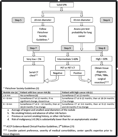
The specific diagnostic pathway can vary depending on individual patient factors, including age, smoking history, and the nodule’s characteristics. It’s important for patients with SPNs to work closely with their healthcare team to determine the most appropriate diagnostic and treatment plan tailored to their unique situation (Figure 1).

Figure 1 The diagnostic algorithm for a patient with a solitary pulmonary nodule (SPN).
The most statistically common cause of solitary pulmonary nodules (SPNs) is benign lung lesions. These benign lesions can have various causes, but among them, some of the most common include:
- Infectious Nodules: Infections such as granulomas (resulting from prior infections like tuberculosis or fungal infections) can lead to SPNs.
- Inflammatory Nodules: Inflammatory conditions like sarcoidosis can cause SPNs.
- Hamartomas: Hamartomas are benign lung tumors made up of a mixture of lung tissues, cartilage, and other elements. They are often found incidentally and are usually non-cancerous.
- Vascular Lesions: Abnormal blood vessels or vascular malformations can sometimes present as SPNs.
- Scar Tissue (Fibrosis): Scarring in the lung tissue due to prior injury or inflammation can appear as a nodule.
It’s important to note that while benign causes are more common statistically, malignant (cancerous) nodules can also be found among SPNs. Therefore, a thorough diagnostic evaluation is essential to determine the cause of an SPN and whether it is benign or potentially cancerous. Medical imaging, biopsies, and pathological examination are key tools in this evaluation process.2
If the biopsy is positive for an NSCLC, what is the behavior?
Non-Small Cell Lung Cancer (NSCLC) is a broad category of lung cancer that includes several subtypes, each with its own behavior and characteristics. The behavior of NSCLC can vary depending on the specific subtype, stage at diagnosis, and other factors. Here are the general behaviors and characteristics of NSCLC:
- Histological Subtypes: NSCLC includes three main histological subtypes: adenocarcinoma, squamous cell carcinoma, and large cell carcinoma. The behavior can differ between these subtypes.
- Staging: The behavior of NSCLC is strongly influenced by the stage at which it is diagnosed. Staging involves assessing the size of the tumor, its extent of spread to lymph nodes and distant organs, and whether it has metastasized.
- Treatment Options: NSCLC is typically treated with a combination of surgery, radiation therapy, chemotherapy, targeted therapy, immunotherapy, or a combination of these approaches. Treatment decisions are tailored to the specific subtype, stage, and individual patient characteristics.
- Prognosis: The prognosis for NSCLC can vary widely. Early-stage NSCLC, when detected and treated before it has spread significantly, generally has a better prognosis than advanced-stage NSCLC. The histological subtype can also impact prognosis.
- Metastasis: NSCLC has the potential to metastasize (spread) to other parts of the body, including distant organs like the brain, bones, or liver. The presence of metastases significantly affects the behavior and treatment approach.
- Survival Rates: Survival rates for NSCLC vary based on factors such as stage and subtype. Early detection and appropriate treatment can improve survival chances.
- Response to Treatment: The behavior of NSCLC can also be influenced by how the cancer responds to treatment. Some tumors may respond well to therapy, while others may be more resistant.
It’s important to note that a positive biopsy for NSCLC is a general diagnosis, and further characterization, including subtype and staging, is necessary to determine the specific behavior and treatment plan for the individual patient. Treatment decisions should be made in consultation with a multidisciplinary team of healthcare professionals, including oncologists and thoracic surgeons, to provide the best possible care tailored to the patient’s unique situation.3
What do molecular biology and genetics contribute in these cases?
Molecular biology and genetics play crucial roles in the assessment and management of lung cancer cases, including those involving solitary pulmonary nodules (SPNs). Here’s how they contribute:
- Risk Assessment: Genetic and molecular factors can influence an individual’s susceptibility to lung cancer. Identifying specific genetic mutations or markers can help assess a person’s risk of developing lung cancer, especially in cases where there is a family history of the disease.
- Subtype Classification: Molecular and genetic analyses can help classify lung cancer into specific subtypes, such as adenocarcinoma, squamous cell carcinoma, or small cell carcinoma. This classification is vital for determining the most appropriate treatment approach.
- Predicting Treatment Response: Molecular testing, such as EGFR (epidermal growth factor receptor) mutation testing, ALK (anaplastic lymphoma kinase) rearrangement analysis, and PD-L1 (programmed death-ligand 1) expression testing, can help predict how well a patient may respond to targeted therapies or immunotherapies. This personalized approach can improve treatment outcomes.
- Treatment Selection: Molecular profiling of lung tumors can guide treatment decisions. For example, if a specific genetic mutation is detected, such as EGFR or ALK alterations, targeted therapies tailored to that mutation may be recommended.
- Monitoring Disease Progression: Monitoring genetic changes in lung cancer over time can provide insights into disease progression and the development of drug resistance. This information allows for adjustments to the treatment plan as needed.
- Prognostic Information: Certain genetic and molecular markers can provide prognostic information, helping healthcare providers and patients understand the likely course of the disease and expected outcomes.
- Clinical Trials: Molecular and genetic information is essential for enrolling patients in clinical trials of novel therapies. These trials often target specific genetic alterations and offer innovative treatment options.
- Early Detection: Research in molecular biology and genetics is ongoing, and there is ongoing exploration of biomarkers that could aid in the early detection of lung cancer or SPNs, potentially allowing for earlier intervention and better outcomes.4
Signs and symptoms
Solitary pulmonary nodules (SPNs) often do not cause specific signs or symptoms on their own. They are typically discovered incidentally during chest imaging studies, such as chest X-rays or CT scans, that are performed for other reasons. However, in some cases, depending on the size and location of the nodule, individuals may experience the following:
- No Symptoms: Most SPNs are asymptomatic, meaning they do not cause any noticeable signs or symptoms.
- Cough: A persistent cough, especially if accompanied by blood-tinged sputum, can sometimes be associated with SPNs.
- Chest Pain: Rarely, SPNs located near the chest wall may cause localized chest pain or discomfort.
- Shortness of Breath: If an SPN obstructs a bronchus or impairs lung function, it may lead to shortness of breath.
- Wheezing: Similar to shortness of breath, wheezing can occur if the nodule affects airway function.
- Recurrent Infections: In cases where an SPN is caused by an underlying infection or inflammation, recurrent respiratory infections might be a symptom.5
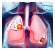
It’s crucial to remember that these symptoms are not exclusive to SPNs, and they can be associated with various other lung conditions as well. If you experience any concerning symptoms or have an SPN detected on imaging, it’s important to consult a healthcare professional for a thorough evaluation and appropriate diagnostic tests to determine the cause and nature of the nodule. Most SPNs are benign, but a proper assessment is essential to rule out malignancy (Figure 2).

Figure 2 Solitary pulmonary nodules (SPNs) often do not cause specific signs or symptoms on their own.
The choice of imaging method to diagnose a solitary pulmonary nodule (SPN)
The choice of imaging method to diagnose a Solitary Pulmonary Nodule (SPN) depends on several factors, including the nature of the nodule, specific clinical needs and the availability of equipment. Here are the main imaging methods used to diagnose SPNs, along with their advantages and disadvantages:
Chest X-ray (Rx of Chest)
Advantages
- Widely available and low cost.
- It can detect SPNs, especially if they are large or dense.
Disadvantages
- Less sensitive for small SPNs or nodules with subtle characteristics.
- Less informative in terms of detailed characteristics of the nodule.
Computed chest tomography (Torax CT)
Advantages
- High sensitivity and resolution, capable of detecting small SPNs and detailed characteristics.
- Provides information about the size, shape, density and location of the nodule.
Disadvantages
- Exposure to radiation, which can be worrisome if multiple scans are carried out.
- Some SPNs may remain indeterminate, which requires additional evaluation.
Chest magnetic resonance (Crax NRN)
Advantages
- It does not use radiation, which makes it safe for certain patients.
- Excellent for evaluating vascular structures and soft tissues.
Disadvantages
- Less sensitive than CT to detect pulmonary SPNs.
- Limited in the evaluation of calcified or small nodules.
Positron emission tomography (PET) with computed chest tomography (PET/CT of Chest)
Advantages
- It can evaluate metabolic activity, which helps to distinguish between benign and malignant nodules.
- It provides information about the extent of the disease.
Disadvantages
- Higher cost and radiation exposure compared to CT alone.
- Less specific to detect small nodules or non-metabolically active SPNs.
There is no single method that is "the best" for all cases of SPNs. The choice depends on the clinical situation and the data obtained from several tests can be combined for a more accurate diagnosis. In most cases, chest CT is the main tool for evaluating SPNs due to its high sensitivity and ability to provide anatomical and morphological details. However, in certain cases, such as when it is necessary to evaluate the extent of the disease or when you want to avoid radiation exposure, other methods can be used. The decision must be made in consultation with the medical and radiological team.6
Treatments
If a neoplastic (cancerous) etiology is ruled out for a solitary pulmonary nodule (SPN), the treatment approach depends on the nature of the nodule and any underlying conditions or symptoms. Here are some possible scenarios and corresponding treatments:
Benign SPN without symptoms
- If the SPN is confirmed to be benign and is not causing any symptoms, it may not require specific treatment.
- Regular follow-up with imaging (usually CT scans) may be recommended to monitor any changes in the nodule over time.
Infectious etiology
- If the SPN is related to an infectious process, such as a fungal infection or tuberculosis, appropriate antimicrobial therapy will be prescribed to treat the underlying infection.
Inflammatory or granulomatous nodules
- In some cases, SPNs may be caused by non-infectious inflammatory conditions or granulomas. Treatment will focus on managing the underlying inflammation or addressing specific conditions if identified.
- Follow-Up and Monitoring:
- Regardless of the etiology, it’s essential to continue monitoring the SPN through periodic imaging to ensure that it remains stable or resolves over time.
Symptomatic relief
- If the patient experiences symptoms related to the SPN, such as pain or cough, treatment may focus on providing symptomatic relief. This could involve pain management or cough medications.
Biopsy or Surgical resection (Rare Cases)
- In very rare cases, a benign SPN may be surgically removed if it causes significant symptoms, grows rapidly, or poses a risk of complications, even if it is non-cancerous.
Radiofrequency ablation (RFA) surgery
The decision to perform radiofrequency ablation (RFA) surgery to treat a peripheral lung nodule near the pleura depends on several factors, including the size and characteristics of the nodule, as well as a comprehensive evaluation by a specialized medical team in pulmonary oncology and interventional radiology. Here are some considerations:
- Nodule Size: RFA is more effective for smaller lung nodules. Smaller nodules typically yield better results with this technique.
- Location: Peripheral lung nodules, close to the pleura, may be suitable candidates for RFA because they are more accessible for the procedure. However, proximity to critical structures such as major blood vessels or the diaphragm can limit its applicability.
- Medical Team Evaluation: A team of healthcare professionals, including interventional radiologists and pulmonary oncologists, should carefully evaluate the nodule, considering factors such as the nodule’s morphology, precise location, and the patient’s overall health before determining if RFA is a suitable option.
- Diagnosis: Confirming the nature of the nodule before any procedure is crucial. This may require a prior biopsy to determine whether the nodule is benign or malignant.
- Treatment Alternatives: Apart from RFA, there are other treatment options for lung nodules, such as surgical resection (surgery to remove the nodule) or microwave ablation. The choice of treatment depends on the comprehensive assessment of the case and the patient’s preferences.
In summary, whether a peripheral lung nodule near the pleura is a candidate for radiofrequency ablation (RFA) depends on multiple factors and should be evaluated by a specialized medical team. The decision will be made based on the safety and efficacy of the procedure in that particular case. Therefore, it’s important to discuss treatment options and potential risks with your medical team to make an informed decision.7
How to make a differential diagnosis between a benign SPN
Distinguishing between a benign solitary pulmonary nodule (SPN) and a malignant (cancerous) SPN is a critical step in the evaluation of lung nodules. Here’s how healthcare professionals typically approach this differential diagnosis:
Clinical history and risk factors
- Review the patient’s medical history, including smoking history, exposure to environmental toxins, and any relevant occupational history.
- Consider the patient’s age and other risk factors for lung cancer.
Imaging studies
- Chest X-ray: Begin with a chest X-ray, which may reveal the presence of an SPN. While less detailed than CT scans, X-rays can provide an initial assessment.
- CT Scan: Perform a high-resolution CT scan of the chest. Characteristics such as size, shape, margin, and density of the nodule can provide valuable information. Malignant nodules may appear irregular, have spiculated margins, and exhibit increased density.
- PET Scan: Positron emission tomography (PET) scans can assess the metabolic activity of the nodule. Cancerous nodules often have increased metabolic activity.
Growth rate
- Monitor the nodule’s growth over time through follow-up imaging studies. Rapid growth may raise suspicion of malignancy, although some benign nodules can also grow.
Calcification patterns
Evaluate the calcification pattern of the nodule seen on CT scans. Benign nodules may exhibit calcification patterns like central, popcorn, or diffuse calcification. Conversely, malignant nodules may show eccentric or stippled calcification or none at all.
Biopsy and pathological evaluation
- If the diagnosis remains uncertain after imaging, perform a biopsy to obtain tissue samples for pathological examination.
- Pathologists can analyze the tissue samples to determine if the nodule is cancerous or benign.
Molecular and genetic testing
- Consider molecular and genetic testing, especially if the diagnosis is unclear or to guide treatment decisions. Specific genetic mutations or markers may indicate malignancy.
Clinical presentation
- Pay attention to the patient’s symptoms, if present. While not definitive, certain symptoms like hemoptysis (coughing up blood) or unexplained weight loss may raise concern for malignancy.
Multidisciplinary evaluation
- Collaborate with a multidisciplinary team, including radiologists, pathologists, pulmonologists, and oncologists, to review all available data and make a comprehensive diagnosis.
Follow-up
- Continue to monitor the patient’s condition through regular follow-up imaging and clinical assessments, especially if the initial diagnosis is indeterminate or if the nodule is not immediately treated.
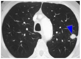
Remember that while certain characteristics and tests can suggest malignancy or benignity, the definitive diagnosis often relies on pathological examination of tissue obtained through biopsy. Differential diagnosis requires a comprehensive approach to ensure accurate and timely management of SPNs (Figures 3 & 4).


Figure 3 & 4 Differential diagnosis requires a comprehensive approach to ensure accurate and timely.
The percentage of lung cancer patients who debut with a solitary pulmonary nodule (SPN) varies depending on the population studied and the diagnostic criteria used. However, it’s important to note that not all SPNs are indicative of lung cancer, and the majority of SPNs are benign. Here are some general observations:
- Incidental Findings: With the increasing use of chest imaging (such as CT scans) for various medical reasons, more SPNs are being incidentally discovered. A significant portion of these SPNs turns out to be benign, unrelated to lung cancer.
- Risk Factors: The likelihood of an SPN being cancerous is influenced by factors such as smoking history, age, and other individual risk factors. High-risk individuals, such as heavy smokers, may have a higher probability of SPNs being associated with lung cancer.
- Diagnostic Workup: The diagnostic workup of an SPN includes imaging, biopsies, and pathological evaluation. Through this process, healthcare professionals can determine whether the SPN is cancerous or benign.
- Histological Types: The type of lung cancer (e.g., adenocarcinoma, squamous cell carcinoma) associated with SPNs can vary. Some types may be more likely to present as SPNs than others.
- Early Stage: Some lung cancers are detected at an early stage when they appear as SPNs. Early-stage lung cancer typically has a better prognosis than advanced-stage cancer.
In summary, while it’s challenging to provide an exact percentage, a significant proportion of lung cancer cases may initially present as SPNs, especially with the increasing use of advanced imaging technology. However, the presence of an SPN does not automatically indicate lung cancer, and a thorough diagnostic evaluation is essential to determine the nature of the nodule and the appropriate course of action.8
Conclusions and final words, for the patient and for the doctor
For the patient
- Awareness and Prevention: Maintaining awareness of lung health and reducing risk factors like smoking and smoke exposure is crucial for preventing lung diseases, including lung cancer.
- Early Detection: Early detection of lung issues, such as lung nodules, can make a significant difference in outcomes. Don’t ignore persistent respiratory symptoms, and participate in screening exams if you’re at risk.
- Communication: Maintain open and honest communication with your medical team. Discuss your concerns, symptoms, and questions. Collaboration with your healthcare team is essential for receiving the best possible care.
- Information and Education: Seek reliable information about your condition and treatment, but avoid unverified online information. Being well-informed can help you make informed decisions and reduce anxiety.
- Emotional Support: Don’t underestimate the value of emotional support. Dealing with cancer and other lung diseases can be challenging, and support from friends, family, or support groups can be invaluable.
For the doctor
- Multidisciplinary Approach: Advocate for a multidisciplinary approach in caring for patients with lung nodules. Collaboration among radiologists, oncologists, surgeons, pathologists, and other specialists is essential for accurate diagnosis and appropriate treatment planning.
- Personalized Treatment: Recognize the importance of personalized medicine. Genetic and molecular testing can help identify the most effective therapies and reduce exposure to unnecessary treatments.
- Empathetic Communication: Empathy and effective communication are crucial. Patients often experience anxiety and fear, and compassionate communication can alleviate their concerns and enhance trust in treatment.
- Promotion of Prevention: Encourage active prevention, such as smoking cessation and reducing exposure to environmental risk factors. Prevention is critical in reducing the burden of lung diseases.
- Continued Research: Support and engage in ongoing clinical and scientific research. Advancing knowledge and treatment of lung diseases, including lung cancer, depends on continuous research efforts.


