Journal of
eISSN: 2376-0060


Case Report Volume 8 Issue 3
1Department of Thoracic Surgery, Ibn Rochd-Casablanca University Hospital, Morocco
2Departement of anatomopatology, Ibn Rochd-Casablanca University Hospital, Morocco
3Hassan 2 University of Casablanca, Morocco
Correspondence: Najat Id El Haj, Department of thoracic surgery Ibn Rochd of Casablanca, 8 Street 1 V/Marjane Polo Casablanca
Received: July 28, 2021 | Published: August 9, 2021
Citation: Haj NE, Hafidi S, Rezouki I, et al. Huge mediastinal bronchogenic cyst revealed by hemoptysis and resected by uniportal VATS: A case report. J Lung Pulm Respir Res. 2021;8(3):104-106. DOI: 10.15406/jlprr.2021.08.00260
Bronchogenic cysts are rare unusual congenital cysts, resulting from abnormal budding of the primary tracheobronchial tube and can be located either in the mediastinum or in the pulmonary parenchyma. They remain asymptomatic in most adults unless they are large to compress adjacent structures or infected.
We report a case of a 43-year-old woman, who presented recurrent episodes of hemoptysis low abundance 4 months ago revealing a huge mediastinal cystic formation, initially taken as a hydatid cyst, the chest CT showed a voluminous cystic formation of 11,4x10, 9x8, 8 cm exerting a mass effect on the posterior mediastinal structures: the superior vena cava, the azygos vein, and the trachea. The diagnosis as a bronchogenic cyst is done after resected by uniportal video-assisted surgery (VATS), and histological confirmation. The patient does not present any complications or reoffending during the follow-up.
The rarity of this entity and even more a rare association of this lesion with symptoms of hemoptysis led to the report of this case. This case illustrates the interest of uniportal VATS for the extirpation of the bronchogenic cyst. We recommend a complete exeresis in most cases to confirm the diagnosis, relieve symptoms, and prevent complications.
Keywords: Bronchogenic cyst, Mediastinum, Hemoptysis, Uniportal VATS
BC, bronchogenic cyst; VATS, uniportal video-assisted surgery
Bronchogenic cysts (BC) are rare birth defects; thought to originate in the primary ventral foregut and occur between day 26 and day 40 of gestation. It is believed that abnormal budding of the primitive tracheobronchial tree occurs in the early stages and develops into a mediastinal bronchogenic cyst, whereas budding that occurs in the later stages becomes an intrapulmonary bronchogenic cyst. Although no particular localization has been identified for intrapulmonary bronchogenic cysts, mediastinal bronchogenic cysts are often located near the bronchi and in the hilar region.1 The first case of the bronchogenic cyst was reported in 1859 by Meyer.2 Since then, many reports have been published. Its incidence accounts for about 5% to 10% of mediastinal masses.3 We recommend through this reported case a complete excision in most cases, why not by uniportal VATS, to confirm the diagnosis, relieve symptoms and prevent complications.
A 43-year-old woman referred to our service for a huge cystic mediastinal formation revealed by several episodes of hemoptysis low abundance 4 months ago, the physical examination was normal, with a PS 0, the chest CT showed a voluminous cystic formation of 11,4x10, 9x8, 8 cm exerting a mass effect on the posterior mediastinal structures: the superior vena cava, the azygos vein and the trachea with partial atelectasis of the right upper lobe Figure 1. The bronchoscopy finds gelatinous secretions that cannot be aspirated completely obstructing the right upper lobe bronchus. Bacteriological samples including finding scolex in bronchial suction fluid, and systematic spur biopsies do not provide additional information. Hydatid serology realized is negative since Morocco is an area endemic for hydatid. Pulmonary function test results showed obstructive ventilatory disorder with a forced expiratory volume in one second of 75%. The decision for surgery was made. Under general anesthesia and after selective intubation, the patient was placed in a left lateral position. A 3 cm incision was made at the level of the 4th intercostal space along the right middle axillary line and a wall protection device was positioned. A 3 cm incision was achieved at the level of the 4th intercostal space along the right medium axillar line and a wall protector was positioned. Visualization of the cyst by thoracoscopy showed that it was very adherent to the superior vena cava, the azygos vein, and the trachea, measuring approximately 11 cm, of regular contours Figure 2. To facilitate surgical dissection of the cystic lesion from the mediastinal structures, fluid aspiration was performed evacuating 200 ml of yellowish and viscous liquid. Once the cyst was empty, complete resection of the cystic lesion was easily completed under uniportal VATS after adhesiolysis dissection of the superior vena cava, azygos vein, and trachea. The procedure was completed with the insertion of a chest tube. The total operative time was 90 minutes and the patient did not need postoperative intensive care. The postoperative (PO) course was uneventful and the patient was discharged home on the 3rd PO day. A control chest x-ray performed after 15 days was normal. Histology examination of the specimen confirmed the diagnosis of the benign bronchogenic cyst with the typical feature of a ciliated columnar epithelial lining Figure 3. The patient is currently in very good general condition and does not present any complications during the long-term follow-up.
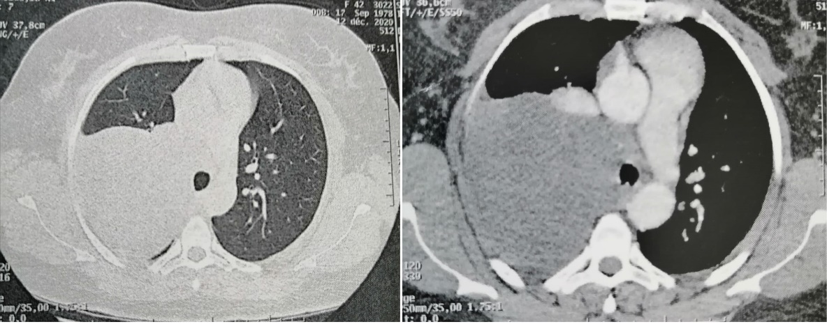
Figure 1 Chest CT axial plan: voluminous cystic formation of 11, 4x10, 9x8, 8 cm exerting a mass effect on the superior vena cava, the azygos vein, and the trachea with partial atelectasis of the right upper lobe.
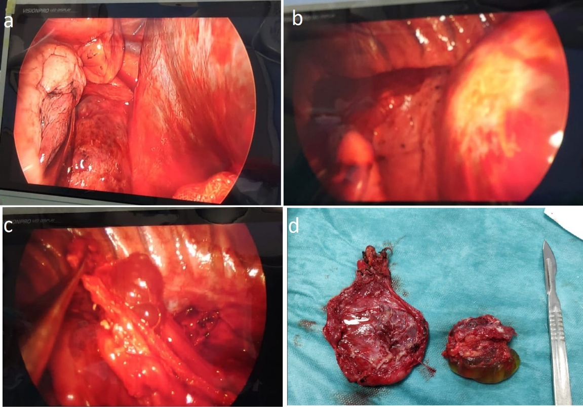
Figure 2 Peroperative image (a,b: the cystic lesion before fluid aspiration, d: the cystic lesion after fluid aspiration, d: specimen).
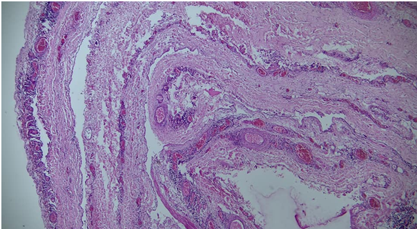
Figure 3 Histopathology with the typical feature of a ciliated columnar epithelial lining.
There are a wide variety of locations where bronchogenic cysts can be found. Most bronchogenic cysts originate from the mediastinum as in our series, with a smaller percentage occurring in the pulmonary parenchyma.
Most BC occur in pediatric patients causing life-threatening compressive symptoms but they are often asymptomatic and incidental radiologic findings in adults.4
Modern imaging techniques such as CT and MRI are useful for precise preoperative diagnosis. The CT scan provides a better definition of the cyst and has eliminated virtually the need for other radiographic studies. Classically bronchogenic cysts are of water density (0-20 Hounsfield units); however, in the presence of infection or those with a variable protein and calcium content, the density may be higher, falling into the solid tissue range and thereby increasing the diagnostic uncertainty. CT images can reveal the relationships between mediastinal bronchogenic cysts and other mediastinal vital structures, and MRI produces T2-weighted images of cyst contents with very high signal intensities.3,5 However, despite advances in diagnostic imaging, a definite diagnosis of a mediastinal bronchogenic cyst is ultimately made based exclusively on the results of a biopsy of the surgically resected tissue specimens. Despite the general absence of clinical symptoms in the majority of patients with mediastinal bronchogenic cysts, severe BC-related complications such as infection, rupture, bleeding, and compression are common. The risk of malignant degeneration such as adenocarcinoma and rhabdomyosarcoma has been reported, and this is one of the reasons why surgical resection is the recommended treatment of choice. In addition, recurrence from incomplete excision has been reported, even after 25 years, and it would seem that complete excision of the cyst wall is desirable whenever possible.6,7
A mediastinal bronchogenic cyst is a benign lesion, and considering recent improvements in thoracoscopic instruments and surgical techniques, the VATS approach is recommended as the first choice of treatment. Minimally invasive techniques in thoracic surgery have been shown to be feasible. The uniportal VATS technique is increasingly used worldwide as a safe thoracic surgical technique for minor and major procedures to treat thoracic and mediastinal pathologies. It should be noted, however, that pleural adhesions are extensive in some cases.8-10
Conversion to open thoracotomy is mainly related to main pleural adhesions and an intact cyst facilitates thoracoscopic dissection along with the layers. A cyst can also open during VATS. Hazelrigg and Associates.11 proposed a deliberate aspiration of the early cyst thoracoscopy to facilitate the handling and preparation of the cyst. In our case, we preferred to suck up the cystic fluid during surgery before extracting the cyst as it was huge. When cysts adhere to vital surgical excision structures may be incomplete, leaving parts of the cyst wall in.11-13 In these cases, the destruction of the mucosal electrocautery lining can help avoid cyst recurrences.14,15 Nevertheless, full excision should always be the primary intention, such as late recurrences, even after 25 years, were described after excision.7 In this study, the removal of the argon beam of the residual cystic epithelium was applied without recurrence after 12 months in one case, but this simple observation does not allow for further conclusions to be made about this management. Incomplete cyst excision is not limited to VATS but was also described after open Surgery.
In this report, a huge mediastinal bronchogenic cyst was compressing the superior vena cava, the azygos vein, and the trachea. The recurrent episodes of hemoptysis were the revealing symptomatology, which was unusual, complete resection was successfully performed by Uniportal VATS, and confirmation was obtained by histopathology. This makes the management of any BC primarily based on complete surgical excision and long-term follow-up for late cyst recurrence.
The authors disclose no conflicts.
None.
The authors disclose no sources of funding for research.

©2021 Haj, et al. This is an open access article distributed under the terms of the, which permits unrestricted use, distribution, and build upon your work non-commercially.