Journal of
eISSN: 2373-4345


Case Report Volume 8 Issue 3
1Department of Oral Radiology, Pontifical Catholic University os Minas Gerais, Brazil
2Postgraduate program of Pontifical Catholic University of Minas Gerais, Brazil
3Department of Oral Radiology, Pontifical Catholic University os Minas Gerais, Brazil
4Department of Oral Pathology, Pontifical Catholic University os Minas Gerais, Brazil
5Department of Oral Pathology, Pontifical Catholic University os Minas Gerais, Brazil
6Department of Oral Radiology, Pontifical Catholic University os Minas Gerais, Brazil
Correspondence: Flavio Ricardo Manzi, Department of Oral Pathology, Pontifical Catholic University of Minas Gerais, Av. Dom Jose Gaspar, 500 / Predio 46 -Coracao Eucarstico-Belo Horizonte/MG, CEP 30535-901, Brazil
Received: September 25, 2017 | Published: September 28, 2017
Citation: Melo NM, Oliveira LJ, Cardoso CAA, et al. Pink spot with an internal resorption: case report. J Dent Health Oral Disord Ther. 2017;8(3):517-519. DOI: 10.15406/jdhodt.2017.08.00284
Introduction: The teeth resorption can occur in the root canal or in the crown. The first one are relatively common and the pink spots in the teeth were first described by Mummery in 1920. Resorption can be classified as internal and external resorption and the internal it is described as a rare occurrence as compared to external.
Case report: this article describes a pink spot that was diagnosed as an internal resorption. The teeth showed with low probability to resist an orthodontic treatment and had to be extracted.
Keywords: pink spot, teeth resorption, unerupted tooth
The teeth resorption can occur in the root canal or in the crown. The root resorptions are relatively common. However, coronal resorptions are rarely seen, especially when they are associated with unerupted permanent teeth.1,2 Moreover tooth resorption can be differentiated into internal and external resorption and occasionally combinations of both can be found on the same tooth.2 Internal resorptions are much less common than external ones.3 Skillen.4 reported the first case of intracoronal resorption in an unerupted tooth in 1941, described it as “intra-follicular caries”. However Kronfeld.5 affirmed that caries couldn’t affect a completely unerupted tooth. O’Neal et al.1 described four theories to explain these preeruptive radiolucencies: apical inflammation of a primary precursor, which affects the permanent successor; dental caries, which has not been proven can develop in an unerupted tooth; developmental abnormality of enamel or dentin, which cause hipoplasia or as an inclusion of uncalcified enamel matrix; internal/external resorption. According to such authors internal resorption is initiated within the pulp cavity whereas external resorption is initiated in the periodontium. Another idiopathic resorption that involves the crown of the teeth was described by Mummary JH.6 in 1920 as a pink spots in teeth. Even though such resorption to be more common in erupted teeth, it can affect unerupted ones. The resorption begins in the pulp and progresses peripherally through the dentin and can involve the cementum and enamel. When the tooth is erupted, the crown discolors and a characteristic pink spot may appear where the pulp is visible through the thinned enamel.3. Typically asymptomatic, the internal resorption associated with unerupted permanent teeth is discovered during radiographic examination obtained to observe disorders of uneruption of permanent teeth or for another reason. In radiographic images, it is presented as well-circumscribed coronal radiolucent área in an unerupted tooth. Histopathologically, evaluation describes numerous capillaries in the pulpal granulation tissue undermining the coronal enamel.2 The pulp tissue in the area of destruction is vascular and exhibits an increased cellularity and collagenization.7 These cases can be treated with nonsurgical root canal therapy (follow-up or pulpectomy) or surgical removal (extraction), which depends on the extension of resorption and on the condition of the tooth, if tooth is erupted or unerupted.8 The following case presents a patient with coronal resorption in an unerupted permanent tooth and the treatment chosen.
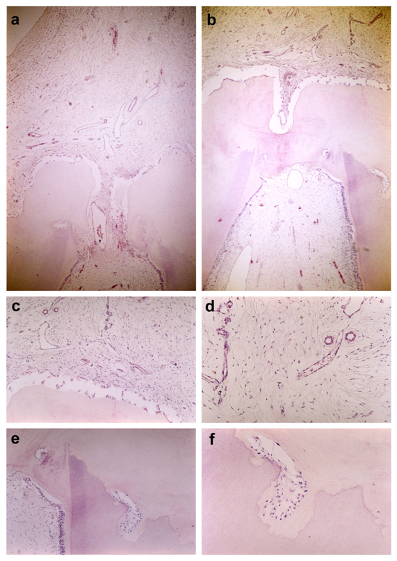
A thirteen-year-old girl was referred to the orthodontic department in a University in Belo Horizonte, Brazil, by her private dentist, to evaluation an absence on the left maxillary canine. Intraoral examination revealed the absence on the left maxillary canine and a swelling in its region (Figure 1), however the mucous was integrity. Radiographs showed the canine unerupted. The tooth presented a wellcircumscribed radiolucent area in the crown. The internal resorption involved the dentin and part of the enamel on this crown (Figure 2). Since the resorption was severe and the enamel of the crown was thinned it was not possible to do the movement by orthodontics treatment. The chosen treatment was tooth removal surgery. During surgery procedure, it was possible to observe that the crown tooth was pink (Figure 3). The tooth was removed and sent to microscopic analysis. The tooth was referred for anatomopathological examination. Histological sections, stained with hematoxylin and eosin (HE), showed that coronary dentin was reabsorbed and replaced by loose fibrous connective tissue. An area of deposition of amorphous acidophilic mineralized material on the reabsorbed dentin region was also observed (Figure 4).

Figure 4 AbsenceHistopathological features. Tooth fragment showing that coronary dentin (D) was reabsorbed and replaced by loose fibrous connective tissue (C). An area of deposition of amorphous acidophilic mineralized material (M) on the reabsorbed dentin region (D) was also observed. The normal dental pulp can also be found (P) (a and b, HE X40; c and e, HE X100; d and f, HE X400).
Teeth resorption is a condition associated with either a physiologic or a pathologic process resulting in a loss of dentin or cementum. Such resorption may occur in root canal or in crown, classified as external or internal. The crown resorption is less common than root resorption.1 Additionally, internal resorption is much less common than external one.3 They can occur in primary or permanent dentition. However internal resorption is rare in permanent teeth.2 Thus the present case joins rare condition of tooth resorption, since it presents intracoronal resorption in an unerupted permanent tooth. Ne et al.2 affirmed that tooth resorption results from injuries to or irritation of the periodontal ligament and/or tooth pulp, as traumatic laxation injuries, orthodontic tooth movement or chronic infections of the pulp or periodontal structures. Eveson & Gibb.3 also indicated various factors as etiology of internal resorption, including vascular changes, acute trauma, restorative procedures, and orthodontic treatment. However, they reported a case, which they considered idiopathic. O’Neal et al.1 defended various theories as etiology of intracoronal resorptions of an unerupted tooth: apical inflammation of a primary precursor affecting the permanent successor; dental caries, which has not been proven can develop in an unerupted tooth; developmental abnormality of enamel or dentin causing hipoplasia or as an inclusion of uncalcified enamel matrix; internal resorption initiating within the pulp cavity or external resorption initiating in the periodontium. Solomon et al.9 described a case where internal resorption were discovered in two teeth after herpes zoster. The authors suggested the hipothesis that idiopathic internal resorption might be related to viral etiology. However Klambani et al.10 defended that etiology of internal resorption is largely unknown. The etiology of present case was considered idiopathic resorption once the patient was not related injuries or other important factor. Gowda.11 in 2015 related two cases about pink teeth with similar variation in the color intensity from region to region and they noted more intense discoloration along cervical region compared to the incised region. Similar to Gowda’s study,11 this present article report a case showed pink discoloration in the dentin and enamel and cementum. Thomas et al.12 described the etiology and pathophysiology mechanisms involved in internal root resorption and they conclude that the prognosis is good for small lesions and when it have a early diagnostic. In our study the diagnostic was latter discovered with an internal resorption involved the dentin and part of the enamel resulting in the extraction. The present report describes a rare case where the internal resorption involved the dentin and part of the enamel on the crown. The treatment resulted in the extraction. After the surgery the patient went to the orthodontic treatment. If they had a early diagnostic, it maybe could prevent the fast progress and the tooth permanence. It is very important the early diagnostic by the dentist to propose the best treatment and prevent the tooth looser.
None.
The authors declare that they have no conflict of interest and the patient provided an informed consent.
None.

©2017 Melo, et al. This is an open access article distributed under the terms of the, which permits unrestricted use, distribution, and build upon your work non-commercially.