Journal of
eISSN: 2373-4345


Research Article Volume 14 Issue 4
São Francisco University, Brazil
Correspondence: Miguel Simão Haddad Filho, Professor of Dentistry at the University of São Francisco, Bragança Paulista SP, Brazil
Received: October 09, 2023 | Published: October 23, 2023
Citation: Haddad Filho MS, Girardello R, Tognetti VM, et al. Evaluation of effectiveness and minimum inhibitory concentration of silver nanoparticles for decontamination of root canal systems. J Dent Health Oral Disord Ther. 2023;14(4):14-19. DOI: 10.15406/jdhodt.2023.14.00604
Endodontics is essential in the process of controlling pain and diseases of the pulp and periapex, thus becoming increasingly innovative, safe and effective. Endodontic treatment has several stages, including the process of canal evolution. This decontamination process is extremely important for the eradication of microorganisms in the SCR and the prevention of reinfection. During the instrumentation it is paramount that constant irrigation occurs to remove inflamed tissues, necrotics and also the biofilm present. The objective of the present study was to analyse the disinfectant capacity of two chemical substances, one of them in different concentrations, o n an aggressive microorganism present in the root canal system. The applied methodology was, in vitro experimental laboratory study, to compare the antimicrobial activity of 1% Sodium Hypochlorite and Silver Nanoparticle at 22 ppm, 70 ppmand 95 ppm, used against the pathogen E. faecalis, which were selected by the bank of microorganisms from the laboratory of Molecular and Clinical Microbiology of the Graduate Program in Health Sciences at Universidade São Francisco (USF), as well as determining the minimum concentration of the silver nanoparticle solution in inhibiting the growth of E. faecalis and P. aeruginosa. Which storage was previously authorized by the Research Ethics Committee of the USF. After data collection, it was possible to conclude from the results that the 1% Sodium Hypochlorite, the Silver Nanoparticle at 70 ppm and 95 ppm, obtained positive results in terms of antimicrobial activity, in comparison with the Silver Nanoparticle at 22 ppm, which obtained a negative result on the microorganism.
Keywords: decontamination, endodontics, root canal
Endodontics is certainly the specialty of dentistry that is becoming increasingly innovative, whether in technological or scientific aspects, making it necessary and indispensable in the treatment of pain. Its meticulous steps require technical and scientific knowledge and manual skills to be executed, and the constant technological evolution allows us to benefit from each advance, in order to constantly improve in this area. Thus within endodontics, which involves subjects such as anatomy, instruments, chemical substances, intracanal medication, among others. Therefore, it is a complex system due to the presence of ramifications, isthmuses, lateral canals, collaterals, recurrent canals, interconduits, dentinal tubules, fins, among other characteristics. These regions are able to promote the formation of biofilm that affect the prognosis of treatment. Generally, the root canal follows the same path as the dentin root. This canal can be curved, straight or sinuous, depending on the internal anatomy of each tooth. Arriving at the end of the root canal, we find the apical foramen, which is an opening at the apex of a root and allows the pulp to be nourished by means of nerves and blood vessels.
When a tooth enters a state of pulpal necrosis, which can occur due to trauma, caries invasion and even the advancement of periodontal disease, the contamination spreads and colonizes the environment. It is an extremely selective region and favors of bacterial proliferation, which involve polysaccharide matrix, where toxic waste occupies the canal and dentinal tubules, with great apical concentration, and also starts to irritate periradicular tissues.1
After preparation, the intracanal medication phase is less and less used, since it is considered that, through the proposal of automated instrumentation, the treatment can be performed in a single session, being control to be done by mechanical action.2
However, regardless of the system chosen, studies show that between 35% to 53% of the root canal walls can remain untouched and thus, exposing the limits of mechanical instruments and showing the importance of chemical substances during root canal preparation and the need for tubule decontamination. That is, it is essential to use disinfectant substances in therapy.
Success of endodontic treatment, the most important is decontamination properly, which takes place by eradicating microorganisms in the SCR and preventing reinfection. During all instrumentation, the canal must have constant irrigation to remove inflamed, necrotic tissue, microorganisms, biofilms and debris from the SCR.
Sodium hypochlorite (NaOCI) is the most widely used irrigant because it has a positive antimicrobial action and the ability to dissolve organic matter, in addition to having a low financial cost. However, it also has some disadvantages, among them significant toxicity when accidentally injected into the periradicular tissue and to some extent affect the mechanical properties of dentin. However, sodium hypochlorite at various concentrations has an excellent effect against the bacteria E. faecalis.3
Substances containing silver nanoparticles (AgNPs) have been increasingly studied as an adjuvant option due the biocompatibility and silver decontamination capacity. Association of NPs with the purpose of disinfection, has specific physicochemical qualities, such as the great ratio of area, surface and mass. Silver ions bind to the cell membrane through the release of NPs, causing the death of microorganisms through the ability that the ions have to damage the bacterial DNA cytoplasm.4
Carreira5 evaluated the ability of the silver nanoparticle solution (AgNPs) used as an irrigant and intracanal medication, as well as controlling microorganisms and neutralizing endotoxins in the root canal. For this, she used 48 standardized human tooth roots and contaminated them for 28 days with E. coli and 21 days with E. faecalis and C. albicans. Later they were divided into 4 groups according to the substance used: G1 –saline solution (control group), G2 –sodium hypochlorite (1%) associated with Endo-PTC cream and calcium hydroxide, G3 –50 ppm silver nanoparticle solution and calcium hydroxide associated with silver nanoparticle solution, and G4 –solution of silver nanoparticle (50 ppm) and five the root canal infected to evaluate the antimicrobial activity. Analyzing the results, it was possible to conclude that sodium hypochlorite and AgNPs solution reduced the microbiota of canal; however, only with the association with calcium hydroxide was there elimination of microorganisms the dentinal tubules and reduction of endotoxins the root canal.
Onoda6 carried out an in vitro study to verify the antimicrobial effect of some endodontic cements added with silver nanoparticles on E. faecalis. He used 3 types of cements: Endofill, Pulp Canal Sealer and AH Plus, later the 3 groups were subdivided into 4 according to the concentration of nanoparticle added to the materials: 0.0%, 0.1%, 0.5% and 1% by weight. Specimens were made and a suspension of E. faecalis was prepared from a 24-hour culture in SBF (Simulated Body Fluid) adjusted in the spectrophotometer. The results showed that cements based on zinc oxide and eugenol used in this study are more effective in eliminating E. faecalis than AH Plus cement, and the minimum addition of 0.1% silver nanoparticle to those cements was effective in improving its bactericidal properties, less so for AH Plus.
Pinto7 evaluated whether the addition of silver nanoparticles to white MTA cement improvement the antimicrobial activity on E. faecalis and avoiding the adhesion of this microorganism to the material. She used the direct contact test and divided the sample into groups according to the materials used, namely: Group A –White MTA, Group B –Gray MTA, Group C – White MTA + 1% AgNps powder and group D – White MTA + 50 ppm AgNps solution and they were kept at 35 ̊C for 72 hours in the E. faecalis suspension. For the comparison of groups considering the variation in number of colony forming units in two periods. There were statistical difference among group C and other groups in the interval T0 to 24 hours, however all brought positive results on the microorganism, already in the interval of 48 at 72, the result was higher for group B when compared to groups C and D. Analyzing the results, the author concluded that the gray and white MTA cements, with or without nanoparticles, showed antimicrobial action on E. faecalis in all periods in the direct contact test, the addition of powdered silver nanoparticle promoted an antimicrobial effect in a shorter time on the pathogens, and in the direct contact test white MTA with silver nanoparticle did not allow adherence after 72 hours in contact with the bacterial suspension.
Wu et al.8 evaluated the antibacterial activity of silver nanoparticles (AgNPs) as an irrigant or medicine against E. faecalis biofilms formed in root dentin surface inoculated E. faecalis for 4 weeks to establish a standard model and then were tested in 2 steps: initially the biofilms were irrigated with a 0.1% AgNP solution, sodium hypochlorite (NaOCl) 2% and sterile saline solution for 2 minutes, respectively. After this, the biofilms were treated with AgNP gel (0.02% and 0.01%) and calcium hydroxide for 7 days. The results showed that irrigation containing 0.1% AgNP solution didn't interrupt the biofilm structure, and the proportion of viable bacteria in the biofilm structures was not different that saline group, but was lower than that of the saline group. Biofilms treated with 0.02% AgNP gel as a drug significantly interrupted the structural integrity of the biofilm and resulted in fewer residual viable E. faecalis cells post treatment compared to 0.01% AgNP gel and groups of calcium hydroxide. Therefore, the authors concluded that the antibiofilm efficacy of AgNPs depends on the mode of application, being used as a medicine it has a greater potential than used as irrigant to eliminate residual bacterial biofilms during root canal preparation.
Almeida et al.,9 compared the effectiveness of solutions of sodium hypochlorite (NaOCl) 1% and 5%, chlorhexidine (CHX) 2%, suspensions of silver nanoparticles (AgNps) 1% and zinc oxide nanoparticles (Np ZnO) 26% versus E. faecalis in 76 single-rooted human teeth mounted in a specific apparatus and sterilized, which 100 μL of an E. faecalis suspension was inserted into the canals, being renewed daily for 7 days. Subsequently, 6 groups were formed (n = 12), according to the irrigating solution used: G1 - 0.85% saline solution (control), G2 - 1% NaOCl, G3 - 5% NaOCl, G4 -2% CHX, G5 - 1% AgNPs suspension and G6 - 26% Np ZnO suspension. The results showed the effectiveness of the 5% NaOCl and 1% Np Ag solutions versus biofilm of E. faecalis, being superior when compared to the 0.85% saline solution. NaOCl 5% reduced 100% of colony forming units compared to the control group, followed by the suspension of AgNps 1% (97.6%), Np ZnO 26% (96.1%), NaOCl 1% (94.1%) and CHX 2% (93.1%). Therefore, 5% NaOCl and 1% Np Ag solutions showed excellent effectiveness against the E. faecalis biofilm established in the root canal.
Samiei et al.,10 were able to evaluate the use of nanoparticles as an antimicrobial agent in endodontic treatments in 15 studies review. AgNPs nanoparticle was the most studied due to its antimicrobial potential. The other nanoparticles studied were bioactive glass and calcium-derived nanoparticles. Observed that the studied nanoparticles present effective and safe antimicrobial activity in the treatment of SCR decontamination. Concluded that the use of nanoparticles presented similar or even better results than conventional irrigants. However, further studies on their toxicity and cellular effects are needed.
Haddad Filho11 conducted a study to compare the disinfectant capacity of four substances used by contact inside SCR consistent with automated proposal for rapid intervention. Fifty human upper lateral incisor teeth were used in the development of the research, extracted and prepared with an automated single file system with externally waterproofed roots. Afterwards, they were contaminated with E. faecalis and centrifugation was performed for tubular invasion. After the samples were divided into 5 groups according to the substance used, namely: photodynamic therapy (PDT), silver nanoparticles (AgNPs), 1% activated sodium hypochlorite (NaOCl), 1% NaOCl and finally the group control with saline solution. Concluded that 1% NaOCl activated by sonic resource was more effective, followed by AgNPs and 1% hypochlorite when contact a short time, finally the PDT did not present significant results, being them similar to that of the control group.
Telles et al.12 carried out a review of the scientific literature in order to verify the scientific knowledge available on the application of nanoparticles in endodontics. Evaluated the antimicrobial activity, prevention versus biofilm formation and elimination, compatibility, resistance to aging, mechanical resistance, dispersion to regions of complex anatomy and tissue regeneration, after searches 50 articles were found addressing the theme and the identified that most of the studies evaluated antimicrobial action and all showed positive results in relation to the incorporation of nanoparticles, while the articles that evaluated the mechanical and structural properties of the materials showed an increase in rigidity, micro hardness and a lower porosity, which is related with increased resistance of these materials. When added to nanoparticles to materials, most studies concluded that the properties were not modified and when there were modifications, they caused a positive effect. Concluded that, an increase in toxicity at high concentrations of nanoparticles can be seen. At the end of the research, they concluded that nanoparticles have been evaluated at the laboratory level and their results are promising in relation to use.
Moradi et al.13 evaluated antimicrobial agents solutions of AgNPs sodium hypochlorite and saline solution as endodontic irrigants of deciduous teeth versus E. faecalis in 36 root canals of deciduous teeth. Specimens were sterilized, soon afterwards they were inoculated with a suspension containing the bacteria, teeth were randomly divided into three groups. Antimicrobial efficacy was evaluated immediately after division into groups by counting colony forming units in brain and heart infusion broth plates. The results showed that 2.5% sodium hypochlorite had the highest antimicrobial efficacy against E. faecalis and showed significant differences compared to the normal saline solution and the AgNps solution at 80ppm, but the latter also showed satisfactory results. Concluded that AgNps solution can also be used.
Rodrigues et al.14 evaluated the antimicrobial action silver nanoparticles in aqueous vehicle, sodium hypochlorite and chlorhexidine on the pathogen E. faecalis. They used bovine dentins that were inoculated in blocks with the biofilm for a period of 21 days, later the blocks were irrigated with a solution of silver nanoparticles at 94 ppm, 2.5% sodium hypochlorite and 2.0% chlorhexidine , for three times: 5, 15 and 30 minutes. The evaluation was performed a confocal laser scanning microscope. The results showed that the silver particle solution was least efficient in eliminating bacteria, however it promoted greater dissolution of the biofilm compared to chlorhexidine. Sodium hypochlorite showed the highest antimicrobial activity and the best ability to dissolve the biofilm. They concluded that the silver nanoparticle irrigant was not as effective against E. faecalis compared to solutions commonly used in endodontic treatment. Sodium hypochlorite was appropriate as an irrigant because it was shown to be effective in breaking up the biofilm and eliminating bacteria in the biofilms and in the dentinal tubules.
Heidar et al.15 investigated and compared the antibacterial and antibiofilm actions of hydroxyapatite nanoparticles and appicability with chlorhexidine digluconate and silver nanoparticles as intracanal drugs in mature biofilm model of E. faecalis in root canals of 68 extracted teeth maxillary central incisors which they were sterilized and infected with E. faecalis and incubated for 28 days under anaerobic conditions to develop a mature biofilm. In 8 teeth were used to monitor the formation and maturation of the biofilm over these days, while the other 60 teeth were divided into two groups of 20 teeth, where one was exposed to 2% chlorhexidine digluconate that interacted with hydroxyapatite nanoparticles and the other second to 0.02% the silver nanoparticle as intracanal medication. In addition, 2 other groups with 10 teeth each were formed. The first group was used as a control positive to verify the bacterial viability throughout the experiment, while the second group was used as a negative control in order to verify the sterility of the procedures. The results showed that both hydroxyapatite nanoparticles functionalized with chlorhexidine digluconate and silver nanoparticles demonstrated effective antimicrobial activity against mature biofilms of E. faecalis.
Marín-Corra et al.16 performed an in vitro study to evaluate the antibacterial effect of a gel preparation containing silver nanoparticles against E. faecalis in 60 root canal extracted single-rooted teeth that were instrumented and later contaminated with E. faecalis. For the antibacterial action, intracanal conduction was performed and 3 groups were formed, the first used 300 ml of gel containing silver nanoparticles, the second 500 ml of gel and the last group was the control, treated with calcium hydroxide. Were incubated at 37°C and a sample was collected daily in 7 days. The results showed that the gel with silver nanoparticles showed effectiveness in antimicrobial action against E. faecalis, the values of minimum inhibitory concentration and minimum bactericidal concentration were 300 g/ml and 900 g/ml, respectively. The authors concluded that the gel containing silver nanoparticles, when used as an intracanal conductive drug in an in vitro model, has an antimicrobial effect. The gel used at 300 ml and 500 ml was equivalent to the action of calcium hydroxide.
Bhandi et al.17 analyzed the effectiveness of silver nanoparticles as root canal irrigants through a bibliographic survey performed searches in PubMed, SCOPUS, Web of Science, and Embase databases, with no restriction regarding publication time. At the end of the searches after going through the inclusion and exclusion criteria, 5 in vitro studies were included in the study and the results showed that silver nanoparticles have an antimicrobial effect to varying degrees. Therefore, it was possible to conclude that silver nanoparticles have the potential to be used as endodontic irrigants, although their effectiveness depends on the particle size and duration of contact.
Neves et al.18 evaluated the antimicrobial action of calcium hydroxide associated with silver nanoparticles on E. faecalis biofilm. In 144 specimens of dentin were inoculated in plates containing culture medium with E. faecalis for biofilm formation. The medications prepared in a 1:1 ratio calcium hydroxide and sterile serum, weighed and added in concentrations of 2.5%, 5% and 10% of silver nanoparticles. 14 days after inoculated, the specimens washed and transferred to a new plate where they placed in biofilm and kept in an oven at 37°C for 2, 7 and 14 days. The samples with biofilm were used as positive control and the specimens without treatment were used negative controls. After experiment time, the specimens washed, shaken, diluted and plated in triplicate in M-Enterococcus. According the findings, it was observed that there was no statistical difference between the measurement of calcium hydroxide with control group in period of 2 and 7 days even after application of concentrations of silver nanoparticles In contrast after 14 days of direct contact with the medication there was a reduction of bacterial biofilm both calcium hydroxide just and application of silver nanoparticles when compared to control group. Concluded that the blend of silver nanoparticles did not contribute to the antimicrobial activity of calcium hydroxide.
Tulu et al.19 evaluated the antibacterial action of silver nanoparticles (AgNPs) mixed with calcium hydroxide or chlorhexidine gel (CHX) against a multispecies biofilm, by confocal laser scanning microscopy (CLSM) and culture-based analysis. Dentin blocks were inoculated with E. faecalis, Streptococcus mutans, Lactobacillus acidophilus and Actinomyceslundii for 1 week. Infected dentin blocks were randomly divided into groups according to medication: group I - saline solution, group II - sodium hydroxide calcium, group III - calcium hydroxide+silver nanoparticles (AgNPs), group IV -2.0% chlorhexidine gel and group V -2.0% chlorhexidine gel+silver nanoparticles (AgNPs). The application times were 1 and 7 days and bacterial samples were collected before and after application of solutions to quantify the bacterial load by staining and confocal laser scanning microscopy.
Results showed that addition of AgNPs to calcium hydroxide increased the antibacterial efficacy at both times of application (1 and 7 days) while addition of AgNPs to CHX was efficient in destruction of microorganisms when compared to all other solutions all application times.
Abadou and Mohamed20 compared the antibacterial action of silver nanoparticles (AgNPs) associated with curcumin paste as intracanal medication, and Ca(OH) calcium hydroxide paste. They used 30 teeth extracted with a single root, these teeth were mechanically prepared, after sterilization, roots were inoculated with E. faecalis for 10 days and separated into 3 groups according to medication used, namely: Group A (AgNPs) , Group B (AgNPs with curcumin), Group C (Ca(OH) paste). The first microbiological samples (S1) were collected from the canal, before inserting intracanal medication. Intracanal medications were kept in root canals in all groups for 7 days and then second microbiological samples (S2) were collected from the canals after medication removal. Results showed that in three groups tested, the highest bacterial count was found in (S1), while lowest bacterial count was found in (S2) with a statistically significant difference between them. The curcumin was superior to the paste of Ca(OH)2. Concluded that AgNPs with curcumin paste had the best antibacterial effect.
Haddad Filho et al.21 objectived in vitro study analyzing the disinfectant capacity of three chemical substances used in endodontic treatment on an aggressive species of microorganism. In carried out experimental laboratory study to compare the antimicrobial potential of 1% Sodium Hypochlorite, 2% Chlorhexidine and 17% Silver Nanoparticle, used in endodontics against the pathogen E. faecalis, selected from the microorganisms culture of the Molecular Microbiology laboratory being included in the sample, 10 isolates of E. faecalis evaluated by the diffusion test. The authors identified that 2% chlorhexidine solution presented best results in relation antimicrobial efficacy, followed by 1% sodium hypochlorite, and finally, 17% silver nanoparticles, which were not able to form growth inhibition against E. faecalis in vitro.
When considering the importance of irrigation and SCR decontamination, as well as the substances used as an alternative to NaOCI, it is justified to carry out a study in order to compare solutions at different concentrations, such as Silver Nanoparticles (AgNPs) at 22 ppm, 70 ppm and 95 ppm, and Sodium Hypochlorite (NaOCI) at 1%, with the purpose of allowing scientific debates as well as contributing to future research and choices to be used by professional dentists. The purpose of the present study to compare two chemical substances, one of them at different concentrations, and to evaluate their antimicrobial capabilities on the aggressive microorganism found in the SCR. The research was carried out in vitro, with the substances NaOCI and AgNPs compared to each other, with the purpose of evaluating results and allowing conclusions.
The microorganisms cultures used were selected of the Microbiology Laboratory (PPGSS Stricto Sensu Graduate Program) in Health Sciences at the University of São Francisco, a clinical isolate of Enterococcus faecalis. The clinical isolate was stored in TSB with 15% glycerol, in a -80°C freezer. Two replicates were performed, on consecutive days, on TSA agar and incubated at 37°C for 24 hours, aerobic conditions. After cultures, the isolate was submitted to the test to evaluate the antimicrobial activity of the tested compounds.
The antimicrobial action of 1% sodium hypochlorite solution was tested against the clinical isolates of E. faecalis. Bacterial inoculate were prepared on the 0.5 McFarland scale (1.5 x 10⁸ Colony Forming Units - CFU), 1 mL of solutions of 1% sodium hypochlorite, 22 ppm silver nanoparticle, 70 ppm silver nanoparticle and 70 ppm silver nanoparticle. silver 95 ppm. The isolate was kept in contact with the solutions 5 minutes at room temperature and then inoculated on surface of a Mueller-Hinton agar plate (Oxoid), with the aid of sterile swab being the plates were incubated at 37°C for 48h. Results were interpreted by visual inspection of bacterial growth on the plate surface.
The bacterial strains were tested in contact with silver nanoparticle solutions 70 ppm, 60 ppm, 50 ppm, 40 ppm, 30 ppm and 20 ppm. The bacterial strains were in contact with the solutions for 5 minutes at room temperature and then transferred to a Mueller-Hinton agar plate using a sterile swab. Plates were incubated at 37C for 24 hours in aerobics. Bacterial growth was assessed by visual inspection after incubation.
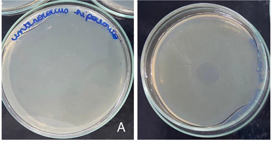
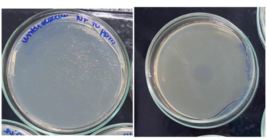
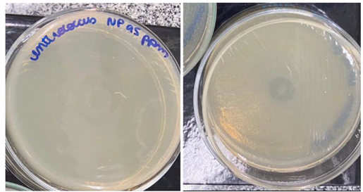
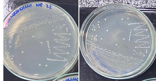
The 1% sodium hypochlorite solution showed antimicrobial activity on the growth of E. faecalis (Figure 1) as well as the AgNPs solutions at concentrations of 70 ppm (Figure 2) and 95 ppm (Figure 3). Therefore, the concentration of AgNPs at a concentration of 22 ppm (Figure 4) was not able to produce any antimicrobial activity on the pathogen (Figure 5 & 6).

Figure 1 1% sodium hypochlorite distributed in a petri dish over E. faecalis. Activity effective antimicrobial action on the bacteria, not showing any residue of the pathogen on the plate. A. Top view of the board. B. Bottom view of the board.

Figure 2 Silver Nanoparticle solution at a concentration of 70 ppm distributed in a petri dish over E. faecalis. The solution showed antimicrobial activity against the pathogen, making it possible to assess that there was no residue on the plate. A. Top view of board . B. Bottom view of the board.

Figure 3 Silver Nanoparticle Solution at a concentration of 95 ppm distributed in a petri dish over E. faecalis. Solution showed antimicrobial activity against the pathogen. A. Top view of the plate. B. Bottom view of the board.

Figure 4 Silver Nanoparticle Solution at a concentration of 22 ppm distributed over E. Faecalis. The solution was not able to show antimicrobial activity against the pathogen, and the bacteria persisted on the plate. A. Top view of board . B. Bottom view of the board.
SCR disinfection depends on the correct technique associated with irrigating substances that have the proper ability to reduce or eliminate bacteria and microorganisms, as a result, several studies have been carried out. It was possible to observe through this in vitro research that the 1% sodium hypochlorite solution showed antimicrobial activity against the pathogen E. faecalis, as well as the silver nanoparticle at concentrations of 70 ppm and 95 ppm, which also obtained a positive result. against the pathogen. Therefore, the silver nanoparticle at 22 ppm was not able to obtain any antimicrobial activity.
It is possible to find similar results in some respects and divergent in others. Samiei et al.10 presented similar results, concluding that the silver nanoparticle is effective and safe in the treatment and decontamination of the SCR, and that the nanoparticles present similar or better results than the conventional irrigants in the present study the tested nanoparticles presented an effectiveness slightly inferior to the compared irrigant. Like Brandi et al., who also concluded through a literature review that silver nanoparticles have potential to be used as endodontic irrigant, but its effectiveness depends on particle size and contact time.
In same way Telles et al.12 concluded that the use of silver nanoparticles has been studied at the laboratory level and that their results are promising in relation to their use. Another similar result was found in the study by Haddad Filho,11 who concluded that activated 1% sodium hypochlorite was more effective against E. faecalis followed by AgNps. Therefore, Carreira5 in an in vitro research, concluded that Sodium Hypochlorite and the AgNP solution significantly reduced the microbiota the canal, but only in association with Calcium Hydroxide was there elimination of microorganisms in depth in the dentinal tubules. Onoda6 states that the minimum addition of 0.1% of Silver Nanoparticle to cements (Endofill, Pulp Canal Sealer) was effective in improving their bactericidal properties, less so for AH Plus. Pinto,7 concludes in his study that the addition of powdered Silver Nanoparticle promoted an antimicrobial effect in a shorter time on the pathogen. Results of in vitro study, Tulu et al.19 concluded that the addition of AgNPs to calcium hydroxide increased its antimicrobial action. Abdou and Mohamed20 concluded that the addition of AgNps was more effective when associated with curcumin paste. In turn, Marim-Corra et al.16 concluded that the association the gel containing silver nanoparticles when used as an intracanal conductive medication in an in vitro model, has an antimicrobial effect, and gel used at 300 ml and 500 ml was equivalent to the action of calcium hydroxide.
On the other hand, we observed divergent results, Neves et al.18 concluded that in the association of AgNps both at concentrations of 2.5%, 5% and 10%, there was no antibacterial activity on the pathogen E. faecalis. Rodrigues et al.14 who, in their in vivo study, concluded that 2.5% sodium hypochlorite is very effective when compared to AgNps at 94 ppm, as well as Moradi et al.13 in which they found that 2.5% sodium hypochlorite had more antimicrobial activity than AgNPs at a concentration of 80 ppm. Haddad Filho et al.21 concluded that 1% sodium hypochlorite showed greater activity than 17% AgNps in their in vitro study, leading to the proposition that the result depends on the concentrations used in the experiment, since in the present study silver nanoparticles in concentrations of 70 ppm and 95 ppm were quite effective, as in the study by Almeida et al.9 who concluded by showing that the 1% AgNps suspension reduced 97.6% of E. faecalis colony-forming units. As stated by Wu et al.8 syringe irrigation 0.1% AgNP solution did not disturbed the biofilm structure, and biofilms treated with 0.02% AgNP gel, as drug, significantly disturbed the structural integrity of the biofilm. Concluded that the antibiofilm efficacy of AgNPs depends on the mode of application, being used medicine it has a greater potential used as irrigant to eliminate residual bacterial biofilms during root canal disinfection.
Silver nanoparticles have been the subject of several researches as an irrigating solution to be considered in endodontics due to their properties such as good conduction and surface plasma resonance, in addition to presenting satisfactory results in relation to the catalytic effect and antimicrobial activity and for covering a larger surface area.22–24 Telles et al,12 through a literature review, identified the silver nanoparticles used as irrigants to be less toxic when compared to other solutions such as Sodium Hypochlorite and Chlorhexidine, in addition to being biocompatible at low concentrations. The results obtained in this research demonstrate that the minimum concentration of Silver nanoparticles to inhibit the growth of bacteria are 70 ppm for the agent Enterococcus faecalis and 60 ppm for Pseudomonas Aeruginosa and such findings are heterogeneous with the findings in the scientific literature.
The 1% sodium hypochlorite solution showed in vitro antimicrobial good activity against the pathogen E. faecalis, as well as the silver nanoparticle at concentrations of 70 ppm and 95 ppm, which also showed good antimicrobial activity. The silver nanoparticle at 22 ppm was not able to produce antimicrobial activity against the pathogen E. faecalis. The minimum concentration required to inhibit the growth of E. faecalis and P. aeruginosa were 70 ppm and 60 ppm, respectively.
None.
The author declares that there are no conflicts of interest.

©2023 Haddad, et al. This is an open access article distributed under the terms of the, which permits unrestricted use, distribution, and build upon your work non-commercially.