Journal of
eISSN: 2373-4345


Case Report Volume 2 Issue 1
1Department of Orthodontics, Aristotle University of Thessaloniki, Greece
2Aristotle University of Thessaloniki, Greece
3Department of Dento-alveolar Surgery, Aristotle University of Thessaloniki, Greece
4Department of Ophthalmology, Medical school, Aristotle University of Thessaloniki, Greece
Correspondence: Georgia Barka, Postgraduate Student, Department of Dentoalveolar Surgery, K Melenikou 9, Thessaloniki, GR54635, Greece, Tel 00306948336960
Received: January 17, 2015 | Published: February 10, 2015
Citation: Ioannidou I, Marathiotis K, Barka G, et al. Dental and orofacial skeletal findings in young male twins with mild Lowe syndrome. J Dent Health Oral Disord Ther. 2015;2(1):8-12. DOI: 10.15406/jdhodt.2015.02.00035
Lowe Syndrome (LS) is a rare genetic disorder involving ocular, renal and central nervous system manifestations. Dental findings have also been described while orofacial skeletal characteristics have not yet attracted considerable attention and estimation. This paper presents the dental findings and the maxillofacial skeletal characteristics evaluated on panoramic and lateral cephalometric radiographies of male twins in mixed dentition. It also provides results of the orthodontic treatment performed to ameliorate the dental and skeletal relationships. Dental and skeletal manifestations of the present male twins suffering from LS as well as the orthodontic treatment approach may enrich the pool of the existing data on this rare syndrome. Moreover, enhanced knowledge may help dentists and health professionals who participate in the overall treatment for these patients to carry out improved treatment plans and provide the best possible quality of life for these patients.
Keywords: lowe syndrome, dental findings, skeletal characteristics, orofacial manifestations, treatment
Lowe syndrome (LS), also reported as oculocerebrorenal syndrome (OCRL), is a rare genetic X-linked recessive disorder. It affects more males than females1,2 and its prevalence was estimated at 1 out of 500.000 individuals with only 190 individuals affected in the year 2000.3,4 LS is caused by a mutation in the OCRL1 gene localized to Xq24-Xq26, resulting in a deficiency of an enzyme called phosphatidylinositol 4,5-biphosphate which regulates the normal activity of the Golgi apparatus.5-7 Several reports described the combination of ocular manifestations (congenital cataract, glaucoma, and nystagmus), manifestations of the central nervous system (mental retardation), and of the kidneys (reduced ammonia production, proteinuria, phosphaturia, and aminoaciduria) for the identification of the LS.4,8-11 Unusual and atypical renal features have also been reported in a mildly affected boy.12 Other significant clinical manifestations include joint hyper mobility, scoliosis, frontal bossing, thin and sparse hair, protruding ears, decreased muscle reflexes and hypotonia.1,13 Oral manifestations associated with LS are more scarcely described and consist of isolated case reports. The reported abnormalities include cases with taurodontism of molars.14,15 crowding and constricted palate, constricted dental arches.4 underdevelopment of the maxilla and mandible, impaction of permanent teeth, gross periodontal disease and severe bone loss. Multiple eruption cysts and odontogenic cyst formation was also reported.1,4 as well as enamel hypoplasia by some.16 and no hypoplastic enamel by others. Delayed tooth eruption was reported in another patient while no delay in the eruption of primary teeth was reported in another. Moreover, in the one case studied with the lateral cephalometric radiography, the analysis showed an increased vertical facial height.10
The present report provides findings on male twins with particular reference to the dental findings and the maxillofacial cephalometric characteristics. It also provides results of the orthodontic treatment performed to ameliorate the dental and skeletal relationships. We believe that reports of different patients suffering of this rare syndrome are useful in order to enhance knowledge about LS, to better define the maxillofacial manifestations related to it, and to approach rehabilitation solutions for the improvement of their quality of life.
Monozygotic twin brothers, 11 years old, were presented to our private clinic referred by their family physician. They requested orthodontic treatment to ameliorate dental arches and facial appearance. Their medical history further includes several typical ocular manifestations. Details of their renal function are unclear, because the patients did not approve further investigation. There was no history of medical condition in the family and according to their mother the twins attended public school and were friendly with their classmates. No behavioral problems were manifested during the first observation. Twins were friendly disposed, intellectual impairment was not noticed but general hypotonia and hyporeflexia were apparent. Both male twins (twin case #1 and twin case #2) at frontal view presented an elongated face, increased lower face height and pale skin. Their ophthalmological examination revealed bilateral congenital cataracts, for which they underwent bilateral lensectomy at the age of 3.5months. Furthermore they presented micro cornea, glaucoma, nystagmus, distichiasis, congenital ptosis of the left eyelid due to partial aplasia of the levatorpalpebrae muscle as well as exotropia of 300 Figure 1 & 2. After the cataract surgery they wore eyeglasses with aphakic correction. Slight prominent forehead and limited height was also observed, sparse hair in the temporal region, as well as deeply set eyes and long eyelashes unusually distant from the eyes. The ears were not protruding but were close to the cranium and low placed. Intra-orally, both twins were at mixed dentition, stage A, presenting malocclusion and relatively good oral health. Panoramic and lateral cephalometric radiographs were procured at the time of presentation. They were taken 6 months before, prior to the extraction of the deciduous molars and the mother was no compliant to take new ones at the present time. Dental impressions were performed and a set of orthodontic study models were obtained for patient’s oral evaluation.


Twin case #1. PM.
Oral examination: At the intra-oral examination, no dental caries were found neither on the maxillary or the mandibular teeth. Hyperplasic gingival inflammation was noticed in the mandibular anterior region (Figure 3).

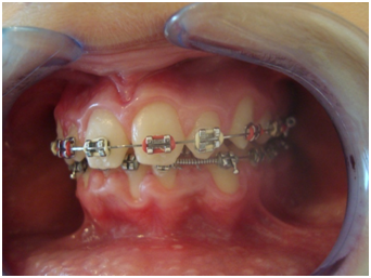
The patient had Class II division 1, the overjet was 5mm and the overbite 4mm. At the maxilla the patient presented remarkably atypical and malformed right and left 1st permanent molars. They were unusually shaped teeth having wedge form. He also presented atypical left and right 2nd primary molars, and hypoplastic and atypical maxillary lateral incisors. The 1st deciduous molar on the right side was extracted and the permanent right canine was under eruption showing the alteration in eruption sequence. The 1st deciduous molar on the left side was also extracted and the lack of space for the eruption of the 1st premolar on the left side was apparent. Atypical morphology of the maxillary central incisors was also found, as well as a diastema between them. The palate was almost normally shaped with mild constriction. In the mandible, both 1st permanent molars and the 2nd deciduous right molar presented atypical morphology. There was rotation of the 1st permanent molar on the left side, the deciduous 1st and 2nd molars on the left side were extracted as well as the deciduous right 1st molar, and there was lack of space for the eruption of the 1st right premolar. The left and the right canine were partially erupted, the anterior teeth presented severe crowding and the mandible showed transverse deficiency (Figure 4).
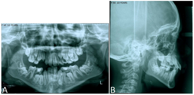
Radiographic examination: Panoramic radiography revealed taurodontism of the maxillary and mandibular 1st and 2nd molars. Delayed eruption of permanent teeth was not seen. On the contrary, the formation of all premolars and the maxillary canines was advanced according to the age. Moreover, the successive development which normally occurs to the posterior teeth was altered as these teeth appeared to have been almost simultaneously developed (Figure 5A). The analysis of the lateral cephalometric radiography according to Ricketts RM.17 (Figure 5B) showed a dolichofacial type of growth, with skeletal Class II malocclusion, high lower facial height, maxillary protrusion and mandibular retrusion. The mandibular incisors were well positioned in relation to the dental plane A-Pog.

Twin case #2. P. K.
Oral examination: At the intra-oral examination of the twin Case #2, no dental caries was found neither on the maxillary or the mandibular teeth and the oral hygiene was good. The patient had a Class III molar relationship left side and Class I right side, the overjet was 2mm, and the overbite 4 mm. A midline deviation toward the left side and a mild transverse deficiency were noticed in the maxilla and the mandible. The study models revealed that the patient presented the same unusual wedge form of the right and left 1st permanent molar and of the 2nd deciduous molar on the left side in the maxilla, like his brother. He also presented hypoplastic and atypical lateral incisors, the left lateral incisor were in crossbite and there was lack of space for the eruption of the 1st premolar on the left side. The deciduous 2nd molar on the right side and the 1st deciduous molar on the left side were extracted; the 1st right permanent molar was rotated, as well as the 1st right premolar which also presented signs of secondary impaction. There was lack of space for the eruption of the 2nd right premolar. Atypical morphology of the maxillary central incisors was also found, as well as a diastema between them. The palate was almost normally shaped with mild constriction. In the mandible, the twin case #2 presented almost the same stage of dental development as his brother. The only difference was noticed in the anterior teeth which presented a mild crowding (Figure 6).
Radiographic examination: Panoramic radiography for the twin #2 revealed the same dental characteristics as his brother. There was taurodontism of the 1st and the 2nd maxillary and mandibular molars, and premature and simultaneous formation of maxillary canines and all premolars according to his age Figure 7A. The analysis of the lateral cephalometric radiography Figure 7B also showed a skeletal Classe II and dolichofacial type of growth, a high lower facial height, good maxillary position and mandibular retrusion. The mandibular incisors were well positioned in relation to the dental plane A-Pog.
Orthodontic treatment for the rehabilitation of the malocclusion was performed in two stages. The first during the mixed dentition with removable appliances and the 2nd stage in permanent dentition with fixed orthodontic appliances. The treatment results obtained for the twin #1 were satisfactory. The mandibular crowding was corrected, the maxillary teeth were aligned, and intercuspation and Class I molar and canine relationship were established (Figure 8A-8C). The treatment results obtained for the twin #2 concerning alignment of maxillary and mandibular teeth, and Class I molar and canine relationship were satisfactory. However, crossbite of the maxillary right premolars remains (Figure 8D-8F). On both twins facial and dental esthetics improved.
LS have been widely studied in the literature and it is of general agreement that for its identification, the combination of ocular and renal manifestations as well as manifestations of the central nervous system is required .1,4,9,10,13. In addition to these relevant features, extra-oral and intra-oral manifestations are described in a limited number of case reports which are characterized by their variability.4,14,15 In the present article, dental and skeletal craniofacial findings of male twins affected by LS are described. It seems to be the second report in the English literature to present cephalometric characteristics of patients with LS. It has been reported that patients with LS have a ‘typical look.13,18,19 They have long face, sparse hair, prominent forehead, and protruding ears. All these features were encountered in our twins with the only exception that the ears were not protruding but were close to the cranium and low placed. Moreover, an unusual feature found in both twins was the blepharoptosis of the left eye. The intra-oral findings concerning the oral hygiene of twin #2 were almost satisfactory. Regarding the twin #1, there were local calculus deposits and gingival hyperplasia in the anterior mandibular region, probably as a result of the crowding of the anterior mandibular incisors. Both patients were able to keep a good level of oral hygiene following our instructions, and accepted positively the recommended regular attendance to the dentist.
The taurodontism of molars is a common feature in patients with LS and it is reported that in this condition, the teeth have normal shaped crown in clinical examination and the taurodontism is only diagnosed radiographically.20,21 Taurodontism of the right and the left 1st permanent maxillary and mandibular molars, as well as of the 2nd deciduous molars was also apparent on both twins radiographically. The particularity is that these teeth in the intra-oral examination presented an unusual shape. They demonstrate a remarkable wedge form and this dental manifestation were not referred in previous reports. Previous studies report an alteration of eruption sequence.13 which is also present in our twin #1 case with regard to his maxillary right teeth, where the canine eruption precedes that one of the 1st and 2nd premolar. However, in our view, the developmental stage of the posterior teeth is the most distinguishing feature observed in our 11 years old twins. Root formation is almost completed in all the posterior teeth and they follow an advanced eruption pattern, as well. Whereas this premature and simultaneous development and eruption of the posterior teeth supports further the absence of a successive pattern of development, it conflicts data generally reported so far about individuals with LS that they present a delayed eruption of their permanent teeth.10,14,16 Our different observation should be taken into consideration as it could be another characteristic oral manifestation of LS which has not previously been sufficiently clearly defined. Moreover, hypoplastic enamel was found in an important number of the twins’ teeth as well as atypical morphology, and this situation has been also referred in other reports.9,16 The transverse deficiency resulting in important crowding is often described in previous reports. The constriction of the maxilla and the mandible was also found in our twins but it was not so relevant to produce crowding except in the mandible of the twin #1.
Mobility of all primary teeth was also reported.9 as well as prolonged retention.10 As mentioned by the twins’ mother, the mobility of the primary canines, 1st and 2nd primary molars in the maxilla and the mandible were the reason that these teeth were extracted. Unfortunately, we did not have the chance to meet the patients before the extraction of their primary teeth in order to evaluate the mobility ourselves. However, early stage mobility could be attributed to the premature development of all the posterior permanent teeth. Cephalometric findings highlighted a convex profile on twins, significant excess in facial height, excess in the lower facial third, posteriorly positioned mandible and anteriorly positioned maxilla for the twin #1 and normally positioned maxilla for the twin #2. Most of these findings are in parallel with the cephalometric findings reported by which was the only report so far in the literature with cephalometric data of an individual with LS Ruellas et al.7 Further reports are required to confirm if these skeletal findings are common characteristics to all the affected individuals with LS and not particular features of our twins.
The manifestation of malocclusion of the male twins is not a particular finding related to them. Malocclusion in individuals with LS has already been described in the limited number of reports4,10 and certainly much more cases are needed to confirm if the malocclusion is part of the LS or it is related to particular patients. The particular condition in the present patients is the rotation and the mesial displacement of the permanent 1st molars in the maxilla and the mandible on both twins as well as the lack of space for the eruption of permanent posterior teeth. These anomalies are obviously the consequence of the mobility and the subsequent extraction of the posterior primary teeth. Therefore, it is very important in the LS cases which present mobility of primary teeth, to better evaluate the developmental stage of the permanent teeth before the extraction of the primary teeth. In cases where the extractions are inevitable, all necessary precautions must be considered in order to avoid additional problems.
The orthodontic treatment performed was satisfactory. Despite the wedge shape of molars and the atypical teeth, Class I molar and canine relationship were obtained, intercuspation as well, and the overjet and the over bite were normalized. Less satisfactory is the transversal harmonization of arches of twin #2. A cross bite of the right upper premolars remains, resulting from the refuse to wear the intermaxillary elastics to achieve better intercuspation. Twin #2 was less cooperative during all treatment and often destroyed appliances. However, both twins and their parents were satisfied for the improvement of facial and dental appearance. The challenge when treating patients with LS to have patient’s cooperation still remains.
In conclusion, patients suffering from LS present a variety of oral manifestations affecting the function of stomatognathic system and the facial esthetics. The present report illustrates oral and skeletal findings of male twins in mixed dentition and the orthodontic approach for rehabilitation of occlusal and dysmorphologic characteristics. Oral and skeletal manifestations related to this rare syndrome need to be better defined with additional clinical and radiographic studies, in order to develop more effective treatment plans and provide best possible quality of life for LS patients.
None.
The integration of this scientific paper was co financed by the project “Scholarships SSF” from the resources of the European program ”Education and Lifelong Learning” of the European Social Fund of the NSRF 2007-2013.
Authors declare that there is no conflict of interest.

©2015 Ioannidou, et al. This is an open access article distributed under the terms of the, which permits unrestricted use, distribution, and build upon your work non-commercially.