Journal of
eISSN: 2373-4345


The extraction of a mandibular incisor constitutes a non-conventional alternative in treating certain anomalies. It is not a standard approach to symmetrically treating most malocclusions, but in some clinical situations the therapeutic aim must be adjusted to individual patient needs, even when this means that achieving final occlusion is not ideal. This treatment option may be indicated in malocclusions with anterior tooth size discrepancy due to narrow maxillary incisors and/or large mandibular incisors. It is contraindicated in malocclusions without anterior discrepancy or with discrepancies caused by large maxillary incisors and/or narrow mandibular incisors. The literature suggests that this method affords improved post-treatment stability compared with premolar extraction. A careful diagnosis, established with the aid of a diagnostic setup, professional skills and clinical experience are instrumental in achieving successful orthodontic results with this treatment option. The deliberate extraction of a mandibular incisor in certain cases allows the orthodontist to improve occlusion and dental esthetics. This article highlights the importance, indication, contraindications, advantages, disadvantages and stability of the results achieved in treatments performed with mandibular incisor extraction. Two clinical cases are presented illustrating the modality of such a treatment.
Keywords: orthodontics, corrective orthodontics, mandibular incisor extraction, Bolton discrepancy, occlusion
Compromised orthodontic treatment may still approach perfection in the denture, providing the result is functional, esthetical in harmony and stable for each case. We believe that a stable, harmonious orthodontic result is dependent upon a healthy occlusion that not only supports its component units but also enhances a healthy periodontium, temporo mandibular joint, and a neuromuscular mechanism. Prior to choose the most favorable treatment option, it is important to analyze treatment goals, stability, the final occlusion to be achieved and the esthetic conditions for each case. In view of this fact, mandibular incisor extraction becomes an alternative treatment for malocclusions that do not fit the conventional forms of extraction since they are more stable in the long term. Although it is not a standard approach to treat malocclusions, but in certain clinical situations, the aim must be adjusted to individual patients needs. The aim of this article is to compile available information in the literature, emphasizing indications, contraindications, advantages and disadvantages, stability of results, limitations, and clinical considerations on the extraction of mandibular incisors as an additional option in correction of malocclusion.
The extraction of healthy teeth has constituted a treatment alternative for over a century. Thus, in 1757 Etienne Bourdet, a disciple of Pierre Fauchard, recommended the removal of the premolars to relieve crowding. Likewise, John Hunter in 1835 extracted the first premolars to allow incisor retrusion in cases of anterior protrusion.1 Hunter reports were published in: "The Natural History of Human Teeth. Edward Angle condemned this practice in the belief that:"...better balance, more harmony and the best possible proportions of the mouth in its multiple relationships require the presence of all teeth and each tooth should occupy a normal position”. Angle also stated in his book: “Treatment of Malocclusion of the Teeth and Fractures of the maxillae’’: “There are cases, however in which extraction is necessary.’’ He then went on to give two reasons for extraction in Class I cases: “First, where the jaws are so small, either naturally or because of arrested development, that the angles of inclination would be too great if all the teeth were placed in line. Second, where extraction is necessary from the requirements of the facial lines, for the development of the arches may be such as to afford an abundance of room for the malposed teeth, and yet the placing of them in the line of occlusion may result in marked dental or labial prominence, and the facial result be more unpleasing than if the teeth had been allowed to remain in malpositions”.2 Angle warned too: “To extract the first upper bicuspid and one of the mandibular incisors, as advocated by some, would only lead to a similar result of less degree besides making impossible the establishment of real harmony between the occlusal planes of the remaining teeth”.2,3 In 1952, Bolton proposed an intermaxillary ratio analysis designed for the purpose of localizing discrepancies in tooth sizes.4,5 Since these ratios form the basis of the present study, a detailed review of their establishment and use will be presented later.
According to Owen,6 the following diagnostic characteristics are usually required for single mandibular incisor extraction:6,7
Diagnostic set-up can demonstrate the amount of interproximal enamel that might be removed from the upper incisors, if that is to be considered. Enamel removal can be distributed among ten maxillary interproximal surfaces (the mesial surfaces of both cuspids and proximal surfaces of the four incisors) to compensate for mandibular incisor extraction. The proximal enamel is usually thick on the mesial surfaces of the cupids’ and the distal surfaces of the central incisors may have only 0.5 mm of enamel. Interproximal enamel reduction is most difficult and hazardous for those teeth which are widest in the cervical region. Excessive reduction of the anterior teeth can cause problems. If all enamel is removed from an interproximal surface, the potential for caries increases substantially, restoration is more difficult, and the teeth may be sensitive to changes in temperature.9,10
The therapeutic extraction of a mandibular incisor may be a solution in selected cases of a tooth-size discrepancy in the canine-incisor region. The orthodontic indications for extracting a mandibular incisor are:
Prevalence of supernumerary teeth in mandibular incisor region is 2% of total supernumerary prevalence, and it is the lowest in the oral cavity. Super numerary teeth in mandibular incisor region may be found in some hereditary syndromes (Gardner Syndrome and acrofacial dysostosis).13,14 Supernumerary eumorphic mandibular incisors may have a familial association. Cassia et al.,15 reported the presence of a supernumerary eumorphic 5th mandibular incisor in a Lebanese family where 4 individuals displayed 5 mandibular incisors with the same shape and size. The hypothesis was a possible autosomal recessive inheritance for this non-syndromic trait. Identifying the supernumerary tooth could be difficult because there is no significant difference between the incisors, or could be easy in case of partial coronal fusion of two incisors. It could be attributed to the decreased available space caused by the presence of supernumerary tooth and the proximity between tooth germs. Because of the inheritance pattern of five mandibular incisors in the studied family, Cassia et al.,15 suggested the involvement of a single gene bearing a recessive mutation.
Congenitally missing mandibular incisors9,16–18
The exact etiology of agenesis of both mandibular central incisors is unknown. Several factors like trauma, radiation, infection, metabolic disorders could be considered. Endo et al.,19 have found that before planning orthodontic treatment on a patient with congenital missing incisors, some factors like reduced mandibular alveolar bone area should be considered.19,20 Some orthodontists like Kokich & Shapiro.,22 and Canutin et al.,1found that congenital absence of both mandibular central incisors is advantageous, as the extraction of mandibular central incisors is considered as the treatment of choice in crowded Class I malocclusion, especially when there is tooth-size discrepancy.21–24
Severe Bolton discrepancy1,10,16
Black25 was one of the first investigators to measure tooth sizes.25 Bolton, analyzed the relationship between the mesiodistal tooth width of maxillary and mandibular teeth. Using the mesiodistal width of 12 teeth, he obtained an overall ratio of 91.3±1.91%; using the six anterior teeth, he obtained an anterior ratio of 77.2±1.65%.26–28 But the average value of the anterior index, 77.2±1.65, cannot be used as a reference point because even the extreme values of this index, 74.5 and 80.4, represent an excellent occlusion.26 A digital caliper was used to measure the casts to the nearest 0.01mm. The width of each tooth was measured from its mesial contact point to its distal contact point at its greatest interproximal distance. Bolton28 anterior (canine to the canine) and overall (first molar to first molar) ratios were calculated with the following formulas:28
For the overall ‘‘12’’ ratio, a significant discrepancy is defined as a ratio below 87.5 or above 95.1, and any ratio below 73.9 or above 80.5 is considered to be a significant discrepancy for the anterior ‘‘6’’ ratio.26 The agenesis of maxillary incisor may lead to this discrepancy. Congenitally, missing maxillary lateral incisors are the second most common dental agenesis, exceeded only by third molars. Selecting an appropriate option for each patient depends on the specific space requirements, tooth size relationship and size and shape of the canines,26 the presence of malocclusion is a primary criteria for making canine substitution the treatment of choice for congenitally missing lateral incisors.
A mandibular tooth-size excess greater than 1.6 mm, is considered significant and can typically be treated by interproximal reduction. Interproximal enamel reduction, or stripping, or air-rotor stripping, or ‘‘slenderization’’, has been advised by Peck & Peck27 as an essential orthodontic treatment ingredient", and has gained popularity in recent. The width of the mandibular incisor crowns can be narrowed by reducing a predetermined amount from each interproximal contact point. The ideal crown shape for reproximation is tapered toward the gingival margin.29–34
Moderate Class III malocclusions with anterior open bite
In patients with a Class III malocclusion, correction is aimed at achieving a Class I key relation and normal overbite and over jet, regardless of the position of the maxilla and mandible. Skeletal change, if needed, is usually limited to the mandible. This means that the relationship of the maxillary first molar to the cranial base is not considered.35 The Anterior Percentage Relation (A.P.R.), which is tied to overbite percentage, expresses the percentage relationship of the maxillary incisor-canine group to the mandibular incisor-canine group36 (Table 1). Removing a single mandibular incisor tooth in a Class III malocclusion is indicated when the mandible is oversize and the A.P.R can be converted into an acceptable figure.37–39 The occlusal area of the mandible is reduced, and by replacing the extracted incisor tooth by adding a first premolar to the anterior segment, the Class III relationship does not need reducing on one side. Quite often this type malocclusion has an open or edge to edge bite.35 The replacement of an incisor tooth with a larger premolar will reduce the size of A.P.R. and the indicated overbite. This could be considered before initiating this type of treatment. An anterior percentage relation of less than 18% after the extraction may produce an open bite.36
|
APR % |
% Overbite |
|
18-Oct |
0 |
|
22 |
15 |
|
30 |
30 |
|
36 |
35 |
|
40 |
50 |
|
55 |
100 |
Table 1 Relationship between APR and overbite. These correlations suggest the limits, as functions of anatomic criteria and functional requirements associated with amount of over-bite, within which mandibular incisor extraction or interproximal stripping are indicated
Ectopic eruption of incisors
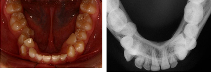
The presence of an ectopic mandibular permanent lateral incisor can result in root resorption and early exfoliation of the deciduous canines and first molars. The diagnosis of such dental anomaly is important for establishing the treatment plan and should be carried out by clinical and radiographic exams. If not treated early, this dental anomaly may develop into partial or complete transposition of permanent canines.40 (Figure 1 & 2).

Class II division 2 with deep overbite
All cases with severe overbite, severe crowding, no tooth size discrepancy anteriorly, anterior tooth size discrepancy due to small mandibular incisors and/or large maxillary incisors, excessive overjet.41
Premolar extraction
Tooth size- arch length discrepancy where premolar extractions are more convenient, and all cases which require maxillary first premolar extraction while canines are in Class I relationship.
Important Class II or Class III skeletal discrepancy42
"Triangular" mandibular incisors15,43
Cases with "triangular" lower incisors and crowding with less than 3mm lack of space should be treated without extractions by stripping the incisors to prevent the reopening of spaces.
Excessive overbite
When the diagnostic setup demonstrates that lower incisor extraction may result in excessive overbite.43–45
High insertion of the mandibular labial frenum10,16
When a high insertion of the mandibular labial frenum may cause gingival recession, but if necessary, surgically lowering the frenum should prevent periodontal complications from occurring at the extraction site.
Gingival recession
Cases where gingival recession may happen in the extraction site in patients with a predisposition to periodontal disease, especially when the roots of the adjacent teeth are not positioned close together.10,46
Brandt & Safirstein et al.,47 have stated the following advantages of mandibular incisor extraction:47
Maintaining the Intercanine width. The extraction space is adjacent to the area of the greatest pre treatment crowding. Although orthodontists continue debating extraction versus non-extraction approaches, post-retention studies indicate a reduction in mandibular Intercanine width over time. In patients with mandibular incisor crowding, both non-extraction and premolar extraction treatment can result in mandibular inter canine width increase. The extraction of a mandibular incisor reduces the dental crowding without expanding the inter canine width
Brandt & Safirstein.,47 have stated the following disadvantages of mandibular incisor extraction:47

The results of a study done by Uribe et al.,49 indicate that 52% of the patients developed an open gingival embrasure after the extraction of a mandibular central or lateral incisor. An open gingival embrasure is a common finding when the distance from the interproximal contact to the crestal bone is more than 5mm.50,51 Faerovig & Zachrisson.,52 reported no cases of black-triangle formation in a sample of patients who had undergone mandibular incisor extractions; they attributed their success to careful selection of patients with little pretreatment crowding, reduction of mesiodistal enamel as needed, and an emphasis on creating optimal axial inclinations of the mandibular incisors.52 The interproximal contact location after treatment was to a certain degree a predictor of the development of an open gingival embrasure. Patients who end treatment with an interproximal contact location at the incisal interproximal third are at greater risk for developing an open gingival embrasure than those who ended with a contact at the gingival third, and this is supported by the study of Kurth & Kokich.,53 They supported too the second reason of open gingival embrasure formation which is root divergence.
Although it may not be possible to eliminate black triangles completely, the risk can be reduced by limiting the distance from the crestal bone tooth contact area. This involves either increasing the bone level in the occlusal direction or moving the contact gingivally. The latter is usually more predictable, and it can be accomplished in one of three ways. First, the root structures must be converged to displace the contact more gingivally, although an extremely low gingival contact will enlarge the incisal embrasure, possibly resulting in uneven incisal edges. Second, the teeth adjacent to the gingival embrasure can be slenderized and the space closed through bodily translation. This option has a potential disadvantage: it may accentuate the anterior Bolton discrepancy created by the mandibular incisor extraction. Third, the incisors adjacent to the extraction site can be built up with composite or veneers. This can be technically difficult, because mandibular incisors tend to be small and often have triangular crowns and roots.
In 1994, Valinoti.,54 suggested that the extraction of a lower incisor54 is less likely to have crowding relapse after retention because the incisor is located closest to the area where the problem is located.16 Reidel.,3 suggested in patients with severely crowded mandibular arches, the removing of one or more mandibular incisor(s) is the only option which allow for increased stability of the mandibular anterior region without retention. In this case, the treatment results would be stable because of the fact that inter canine width is decreased, and the mandibular incisors are not protruded.5 Reidel.,3 wrote: “The extraction of two mandibular incisors may satisfy the requirements of maintaining arch form without expansion of inter canine width’’. With non-extraction or premolar extraction therapy, the original inter canine width usually must be increased in order to gain adequate alignment and arch form, a strategy that might be result in a more favorable result.3 Reidel et al.,3 suggested extracting a mandibular incisor can provide greater stability in the anterior area in the absence of permanent retention. They evaluated the pretreatment, post-treatment, and 10-year post-retention records of 42 patients. Each patient had one or two mandibular incisors extracted before complete orthodontic treatment. They compared the overall stability of patients treated with premolar extractions and those treated with extraction of one mandibular incisor. They found more acceptable mandibular incisor alignment in those patients treated with a single incisor extraction at post-retention (29%). The premolar extraction cases demonstrated 70% unacceptable incisor alignment at post-retention. Canut also found better stability in patients who had one mandibular incisor extracted when compared with patients requiring premolar extraction. He evaluated the pretreatment, post-treatment, and 5 to 8 years post-retention records of 26 patients.1
Gilmore & Little.,55 evaluated the incisor dimensions and stability by evaluating the post-retention records of 134 patients who had undergone orthodontic treatment at the University of Washington. They found a weak tendency for narrower incisors to be associated with improved alignment. However, their results indicated that narrower mesiodistal widths of the mandibular incisor crowns did not guarantee better long-term stability of mandibular incisor alignment.55 Salzmann et al.,2 suggested that extracting a mandibular incisor would result in an excessive overbite. Kokich & Shapiro.,22 believe that the problem of increased overbite can be avoided by carefully evaluating the complete diagnostic records in selecting suitable patient for this treatment plan. They also believe that in case selection, the intentional extraction of a mandibular incisor can simplify orthodontic mechanics and enhance both occlusal and cosmetic results of treatment. Success in treatment depends upon patient selection and a mandatory “diagnostic wax set up” before making the extraction decision.22,56–58
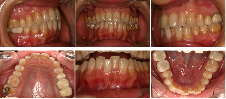
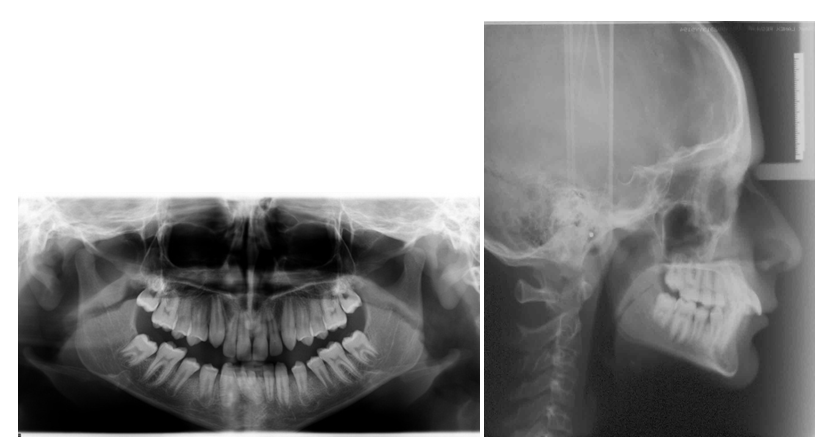
Case 1 Extraction of a mandibular incisor (Figure 4–9)
Case 2 Missing one Mandibular incisor (Figure 10–15)


Mandibular incisor extraction can be an effective treatment option for malocclusion with a Bolton discrepancy. The incisor extraction decision must be supported by a large inter canine width, relatively minor crowding, some mandibular anterior tooth size excess, and normal rather than triangular incisor shape.50 Several factors must be considered before making the final treatment decision. Evaluation of a diagnostic wax set-up will allow predicting the success of the proposed treatment plan.59–62
This treatment option may cause some difficulties or limitations in orthodontic treatment: obtaining group function, possibility of spaces reopening, loss of gingival papilla, impact on the midline, increased over jet and overbite, formation of open gingival embrasures or black triangles. It is difficult to predict the risk of this phenomenon, but it may be an important esthetic consideration, especially in older patients. Although the indications for this type of extraction decision are relatively rare, the possibility of incisor extraction should be an option of every orthodontist’s portfolio of treatment techniques. If properly indicated, mandibular incisor extraction can contribute to the treatment of certain malocclusions and achieve excellence in orthodontic treatment results.
None.
The author declares that there are no conflicts of interest.
None.

© . This is an open access article distributed under the terms of the, which permits unrestricted use, distribution, and build upon your work non-commercially.