Journal of
eISSN: 2373-4345


In recent years dentists have learned that the metal fillings in the mouth act much like metal does outside the mouth. Exposed to hot and cold, such as hot coffee or ice cream, the metal fillings in the teeth expand and contract. As they shift, they can actually weaken the teeth they were meant to protect. Often, the expansion and contraction leads to the entire tooth cracking. As the fillings expand it can also leave a small opening where harmful bacteria can enter and become trapped, leading to further decay of the tooth. Fracture lines in teeth create further avenues for decay to occur. Rarely, do any doctors remove silver fillings without finding additional decay underneath. An Inlay or Onlay is a much more conservative restoration for the tooth than a metal filling or even a crown. While traditional fillings can reduce tooth strength up to 50%, inlays and Onlays made of high strength porcelain or composite systems (ADORO), can actually increase tooth strength by up to 75%, lasting 10 to 30years. An Inlay is similar to a filling and lies inside the cusp tips of the tooth (as we explain to our patients for better understanding, is a filling which is constructed at the laboratory). Onlays are used for large restorations. They restore the area inside the cusp and extend over one or more sides of the tooth. Basically, it covers one or more cusps of a tooth. Onlays are indicated in situations where a substantial reconstruction is required. However, more of the tooth’s structure can be conserved compared to the placement of a crown.
Keywords: inlay, onlay, cavities, fillings, bonding, impression, ceramic, restoration, micro leakage
MOL, mesial occlusal distal; UDMA, urethane dimethacrylate
Inlays and Onlays are forms of indirect restoration used when a molar or premolar is too damaged to support a basic filling, but not so severely that it needs a crown. From our clinical experience, it seems that we can apply Onlays in “extreme” cases, for example; in a molar with 2 or even 3 cusps missing. Inlays and Onlays are prepared outside the patient’s mouth, then are cemented or bonded to the tooth. The inlay or Onlay fits into the prepared tooth much like a puzzle piece and are intended to rebuild a large area of chewing surface of a tooth, whereas fillings are direct restorations designed to fill a small hole in tooth enamel. Inlays and Onlays are not as extensive as crowns which cover most of the tooth. An Inlay is placed on the chewing surface between the cusps of the tooth, while an Onlay covers one or more cusps. Onlays, which are sometimes called partial crowns, may be used if more than half of the biting surface of the tooth is decayed or otherwise in need of repair.
Preparing and placing inlays and Onlays is a multistep process, it involves:
If a dentist has the appropriate equipment (CAD/CAM), this can be done in a single visit to the dentist. A temporary inlay or Onlay is placed on the prepared tooth while a patient waits for the finished restoration to return from a dental laboratory. Materials such as gold, composite resin or ceramics may be used to create inlays or Onlays. Which material is chosen may be influenced by aesthetic appeal, strength, durability and cost. The material used plays a major role in determine how long those restorations will last, as some substances are tougher and better tolerated than others. Other factors that influence the longevity of an inlay/Onlay include the strength of the tooth that is treated, the amount of chewing that occurs on the restorations and a patient’s willingness to maintain oral hygiene and to have regular examinations. As I previously referred, the clinical applications of inlays and Onlays are really impressive because with a minimal invasive restoration, we succeed a perfect bio-esthetical result.
The main indications for constructing a ceramic or composite inlay or Onlay are1
In cases which the preparation of the teeth gives the appropriate support or a sufficient amount of enamel for a successful bonding, the construction of an Onlay in cases we have excessive loss of tooth tissues is possible. The presence of enamel is important because the durability of the adhesive interface with enamel is very predictable.
The advantage of ceramic inlays and Onlays are:
The disadvantage of ceramic inlays/Onlays is:
Case 1
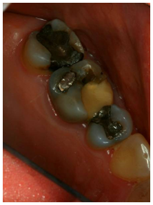
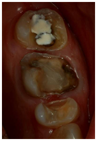
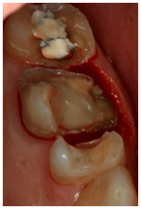
A 30year old female patient presented with some micro fractures in the two upper left premolars with mesial occlusal distal (MOL) dental amalgams. There were no symptoms. After options of another direct composite filling and crowns were discussed we decided to restore the two teeth with two.2 suggest that various studies show that ceramic inlays perform better in females than in males. The same authors also support that ceramic inlays survive better in vital molar teeth and that premolar ceramic inlays have more longevity than molar ceramic inlays. We started by removing the amalgam fillings and then we start preparing the teeth.
To prepare the tooth for an inlay or Onlay, we must have in mind some principles:
Having in mind all these principles, we start preparing the tooth. After we finish the preparations, we are ready to take impressions.
In our case before we apply flow able composite for sealing the undercuts we use an opaque composite material o cover the dyshronic dentin caused from the amalgam filling. For the preparation, in our centre we use the set for universal preparations which are manufactured by Komed. The next stage is to take an excellent impression, and an excellent impression is the one that can be read by the technician. In our centre we use the technique of double impression. If it is necessary (when the limits of the preparation are in the same level with the gums or lower) we put two cords #0, #1. The #0 cord stays in the gingival crevice during the impression. When we have to take the impression for a single Onlay we use a triple tray and a combination of putty and light body vinylpolysilotane in the double impression technique. After we check that the impression is correct we choose a colour and apply a temporary filling material. If it is necessary we send the laboratory some photographs. The last twoyears in our centre we have “abonded” the ceramic Onlays and in co-operation with the lab we use a new system. The new SR-ADORO3 system is a composite system that offers several advantages over hybrid composite materials as regard to new handling, plaque resistance and surface finish. The advantageous properties of SR ADORO can be attributed to the high proportions of inorganic fillers in the nanoscale range. Furthermore, the matrix is based on a urethane dimethacrylate (UDMA) which has also been newly developed and which is characterized by its toughness which is higher than of its predecessors or the frequency used Bls GMA. The material demonstrates colour stability as well as outstanding enamel like structure and material opalescence. The aesthetic appearance that can be achieved is impressive. When the Onlay comes back from the lab we check whether it fits perfectly in the cast. Then we check the thickness of the restoration. We want to have 2mm thickness especially in stress bearing areas. If we have less there is a possibility for the restoration to fracture. If the thickness is not correct then the lab will inform us to prepare deeper. If everything is fine we move to the bonding procedure which is again a very important stage for the success of the restoration.
It is generally recommended that the use of rubber dam isolation provides greater restoration predictability than cotton roll isolation.2 suggested that the overall restoration longevity reported in long term studies does not appear to be adversely affected by the type of isolation technique. In our centre we first remove the temporary filling and clean the tooth by applying air-blasting. Then we try the Onlay in the cavity and check the fit, the colour and the contact points. At this stage we don’t check the bite. Because we deal with Onlays made with the new ADORO system the bonding procedure is easy. We etch the tooth; the enamel for 30seconds and the dentin for 15seconds. We apply the EXCITE DSC (IVOCLAR)4 bonding agent to both the tooth and the restoration. We don’t photo polymerize the adhesive. We then apply VARIOLINK II to the cavity and we locate he restoration to the cavity. We remove the excess cement, we apply a gel called liquid strip to prevent the polymerization from the oxygen and then we polymerize each surface for 40seconds. We check the bite and if necessary we remove some of the material with smooth diamond burs. In the interproximal surface we use polishing strips. Finally we polish the restoration with silicon polishers (ASTROPOL) (Figure 1–3).


Case 2
A 55year old woman came to our centre. She was not happy with the restorations to teeth 25, 26, 27. We proposed her to have Onlays and she agreed. We started to remove the old fillings. In tooth 26 after we removed the old fillings and cleaned the cavity from the decay the sound tooth structure was 0.75 mm below the gum. We decided to remove the excessive gum tissue and some bone if necessary so as the sound tooth structure would be at least at the same level with the gum. Concerning the biologic width we removed the gum tissue with Er-YAG laser (2940nn wavelength). In five days we saw the patient again. Postoperative healing was excellent so we took impressions and in two days the lab sent back the restorations. We have chosen a lighter colour because the patient informed she was planning to have aesthetic restorations in her other teeth (Figure 4–9).


Case 3
This patient came to the centre with a missing amalgam filling. We just cleaned the decay we took an impression and the next day the lab sent us the Onlay which had a perfect fit. After we clean the teeth we apply a caries indicator so as we are 100% sure that all the decayed area has been removed. We took impressions and in two days we fitted the restoration (Figure 10–13).
As we saw before, ceramic inlays/Onlays are very promising restorations regarding the aesthetic factor. Briefly the advantages are:-
I am next going to refer to some surveys about Inlays and Onlays which support the use of these restorations in the daily clinical practice.
Barghi & Berry5 show in their study that porcelain overlays with supragingival margins entirely on enamel that rely primarily or entirely on bonding for their retention, can provide excellent aesthetics, good function and perhaps long term durability if properly designed, fabricated and bonded. Porcelain overlays fabricated from high leucite content porcelain, bonded to sound enamel and dentin with a dual-cure luting resin, and a fourth generation dentinal adhesive provide satisfactory clinical results and high patient satisfaction.
Frankenberger et al.6,7 show that IPS Empress Inlays and Onlays, exhibited satisfactory clinical outcomes over a 12 year clinical period. Restorations luted with dual-cured resin composite, revealed significantly fewer bulk fractures. Guess et al.8 found that all ceramic materials IPS e-max and PRO CAD, seem to be indicated for partial coverage restorations on molars. Prakki et al.9 compared various cements and they concluded that Variolink II exhibited the least weight loss and roughness increase.
Van Dijken et al.10,11 evaluated the marginal breakdown of fired porcelain inlays in vivo by which were luted with either a dual-cured resin composite or a glass polyalkenate (ionomer) cement by scanning electron microscopy. They concluded that the overall marginal quality was significantly better for the inlays luted with the resin composite both at baseline and after one year. Meyer et al.12 supported that when posterior teeth are weakened, owing to the need for wide cavity preparations, the success of direct resin based composite is compromised. In these clinical situations, ceramic inlays/Onlays can be used to achieve aesthetic, durable and biologically compatible posterior restorations. Kramer et al.13 found IPS Empress Inlays and Onlays bonded with syntac classic were found to have a 92% survival rate after eightyears of clinical service.
Kramer et al13 evaluated the effect of two different adhesive resins composite combinations for luting of IPS Empress inlays. Syntac/Variolink II, IBS (3M ESPE). They concluded that the luting of ceramic inlays, no difference between the two luting systems was detectable. The overall failure rate after 4years was 4%. Barone et al.14 found that composite inlays demonstrated a very high success rate (97.4%) after threeyears. Neither the size of the restorations or the tooth type significantly affected the clinical outcome of the restorations. Manhart j et al.15 showed that posterior tooth coloured inlays exhibited a success rate of 100% for ceramic inlays and 90% for composite inlays, even if placed by relatively inexperienced bit supervised student operators.
Van Dijken et al10,11 found that the success rate of the dentin enamel bonded ceramic coverage’s, reduces the need for a traditional full-coverage therapy and/or post or pin (s) and core placement. This technique showed many clinical advantages such as less destruction of healthy tissue, and avoidance of endodontic treatment and/or deep cervical placement of restoration margins. Frankenberger et al.6,7 found that etch and rinse adhesives combined with conventional luting resin composites reveal still the best prognosis for adhesive luting of glass ceramic inlays. Stappert et al.16 concluded that since the majority of IPS e-max press and ProCad restorations survived loads within the range of physiological mastication forces, both materials appeared to be suitable for the predictable use of posterior partial crowns.
We must also have in mind that traditional fillings can reduce the strength of a natural tooth by up to 50%. As an alternative, inlays and Onlays, being bonded directly onto the tooth using special high strength resins, can actually increase the strength of a tooth by up to 75%. These restorations are already an excellent choice for the clinicians and in combination with the entrance in the dental market, more technologically developed systems with more reinforced materials there is a solid optimism for the future application of these restorations to the daily clinical practice.
None.
None.
Authors declare that there is no conflict of interest.

© . This is an open access article distributed under the terms of the, which permits unrestricted use, distribution, and build upon your work non-commercially.