Journal of
eISSN: 2373-4396


Case Report Volume 11 Issue 6
1Department of Cardiology, Kosuyolu Kartal Training and Research Hopital, Turkey
2Department of Cardiology, Hitit University, Faculty of Medicine, Turkey
3Department of Cardiology, Ümraniye Training and Research Hopital, Turkey
4Department of Cardiology, Ümraniye Training and Research Hopital, Turkey
Correspondence: Macit Kalcik MD, Department of Cardiology, Hitit University, Faculty of Medicine, Turkey, Tel (90)536 4921788, Fax (90)3645117887
Received: October 22, 2019 | Published: November 15, 2018
Citation: Güner A, Kalç?k M, Bayam E, et al. Visualisation of an interesting vegetation on a mechanical prosthetic mitral valve in an intensive care unit patient with infective endocarditis. J Cardiol Curr Res. 2018;11(6):229-231. DOI: 10.15406/jccr.2018.11.00405
A 48-year old man was consulted to cardiology for 39°C fever and ongoing dyspnea for 2 days during his hospitalization in the intensive care unit. He had undergone mechanical mitral valve replacement 3 years earlier. His history included a dental procedure without antibiotic prophylaxis 15 days ago. The chest radiogram was unremarkable. Transthorasic echocardiography revealed a mass on the mechanical mitral prosthesis and increased transvalvuler gradients with decreased valve area. Subsequently, transesophageal echocardiography (TEE) was performed which showed a large obstructive mass on the prosthetic valve, protruding into the left ventricular cavity during diastole. It was observed that, the cross sectional images of the mass was resembling various animal shapes in different multiplane TEE views
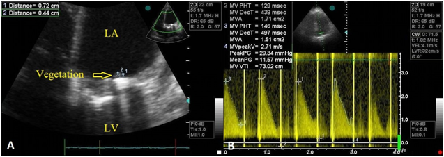
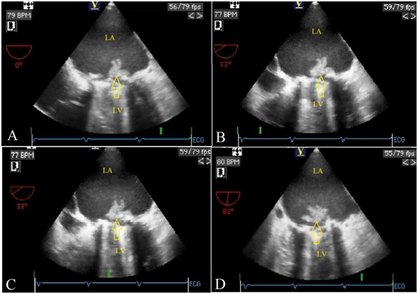
A 48-year old man was consulted to cardiology for 39°C fever and ongoing dyspnea for 2 days during his hospitalization in the intensive care unit. He had undergone mechanical mitral valve replacement 3 years earlier. His history included a dental procedure without antibiotic prophylaxis 15 days ago. The chest radiogram was unremarkable. Transthorasic echocardiography revealed a mass on the mechanical mitral prosthesis and increased transvalvuler gradients with decreased valve area (Figure 1) Subsequently, transesophageal echocardiography (TEE) was performed which showed a large obstructive mass on the prosthetic valve, protruding into the left ventricular cavity during diastole. It was observed that, the cross sectional images of the mass was resembling various animal shapes in different multiplane TEE views. A rabbit like image was obtained from midesophageal transverse plane (0 degrees) (Figure 2A) (Video 1). Whereas on 33 degrees, the same image resembled to a mouse head (Figure 2B) (Video 2). Interestingly, after clockwise rotation of the probe shaft at the same degree the mouse image turned into a cow’s head with its horns (Figure 2C). Finally, with the transducer array at 82 degrees, the vegetation looked like a flapping pigeon (Figure 2D) (Video 3). Consequently the patient underwent redo valve surgery and was discharged after an uneventful postoperative period. Since two-dimensional TEE provides cross sectional analysis of cardiac strutures, masses with irregular borders can be viewed as different shaped images in different TEE views.1 We considered this case valuable as 4 different animal-like images were depicted in the same patient.

Figure 1 Transthorasic echocardiography revealing a mass on the mechanical mitral prosthesis. (A) Increased transvalvuler gradients with decreased valve area, obtained by continuous wave Doppler imaging. (B) (LA, Left atrium; LV, Left ventricle).

Figure 2 Two-dimensional transesophageal echocardiography showing a rabbit like image , obtained from midesophageal transverse plane (0 degrees). (A) An image resembling to a mouse head with big ears, eyes and long nose at 33 degrees. (B) Clockwise rotation at 33 degrees resulted in a cow’s head image with its horns. (C) With the transducer array at 82 degrees, a pigeon image was obtained with it’s wings, tail and opened beak. (D) (LA, Left atrium; LV, Left ventricle).

Video 1 Two-dimensional transesophageal echocardiography showing a rabbit like image, obtained from midesophageal transverse plane (0 degrees) (LA: Left atrium, LV: Left ventricle).
No additional data
All of the authors contributed planning, conduct, and reporting of the work . All contributors are responsible for the overall content as guarantors.
No funding.
We have nothing to declare
None.
All of the authors have no conflict of interest.

©2018 Güner, et al. This is an open access article distributed under the terms of the, which permits unrestricted use, distribution, and build upon your work non-commercially.