Journal of
eISSN: 2373-4396


Case Report Volume 11 Issue 6
1Area of Health Sciences, Autonomous University of Zacatecas, Mexico
2Chair, Department of Cardiology, National Institute of Cardiology, Mexico
3Ignacio Morones School of Medicine, Mexico
Correspondence: Juan Manuel Cortes Ramírez, Department of Health Science, University of Zacatecas, Mexico
Received: October 25, 2018 | Published: November 5, 2018
Citation: Ramírez JMC, Edith CV, Torre JMJC. Turner syndrome case presentation. J Cardiol Curr Res. 2018;11(6):221-223. DOI: 10.15406/jccr.2018.11.00403
Humans Have 46chromosomes. Two of them sex: the X and Y. X the two women (one from the father and one from the mother). Males have one X (the mother) and one Y (his father). In the early stages of cell division, division Makes a wrong part or all of the X chromosome Circumstances That influence, Most Often lost is unknown chromosome is lost to the father. The diagnosis of ST requires a phenotypic Characteristic with full or partial absence of one X chromosome, as phone line or regulate as to mosaicism. The ST 1/5 1/2 prevalence has 000 000 female births. It is characterized by gonadal dysgenesis and short stature. At birth with lymphedema, redundant skin and webbed neck. In childhood stunting and pubertal development of subsequently absence and primary infertility is detected, Accompanied by epicanto, wide neck, short with pterygium, low-set hair, broad chest, shield, breast hypertelorism, cube valgus, hands and feet lymphedema. Normal psychomotor development and IQ. Structural abnormalities that May be present: Kidney. Eyepieces of middle ear disease. A craniofacial level. In skin: An orthopedic level. It is associated with inflammatory bowel disease. Autoimmune thyroiditis; Basedow Graves disease, especially X. With isochromosome Obesity, Glucose intolerance, type 2 diabetes, hypertriglyceridemia, and insulin resistance. Frequently cardiovascular malformations in 45, systemic arterial hypertension X. After the suspected or confirmed, apply to protocol detection, monitoring and treatment of various comorbidities, for an adequate quality of life.
Keywords: Turner syndrome, short stature, chromosome X
Humans have 46 chromosomes in the cells. Two of them are called sex: the X and Y. Women have two X chromosomes (one from the father and one from the mother). Men have an X chromosome (the mother) and another Y (his father). In the early stages of cell division giving rise to an embryo, an erroneous division makes part or all of the X chromosome is lost if the pregnancy continues, the child will have Turner syndrome (TS). This does not occur in children who have only one X chromosome and if needed, could not live.It is unknown what circumstances influence is abnormal division to occur, most often chromosome lost to the father. The cause is not known. (One) ST diagnosis requires combining a certain phenotypic characteristics with a total or partial absence of an X chromosome, as well as regulating cell line or mosaicism. It is one of the most common monosomy, prevalence of 1/2 000 1/5 000 female births.1,2
It is characterized by short stature and gonadal dysgenesis. One-third are recognized at birth by lymphedema, redundant skin or webbed neck. Another third, in childhood, stunting and the remaining third when not present pubertal development or primary infertility. Among its highlights dysmorphic epicanto, pinnae rotated back, wide, short neck with pterygium, low hairline, broad chest, shield, separated breasts, cubitus valgus, hands and feet with lymphedema, narrow and concave nails. Psychomotor development and IQ are normal (two) structural abnormalities that may be present: kidney 30-40%. 50-85% have middle ear disease: recurrent suppurative otitis media, serous otitis media, chronic suppurative otitis with perforation, conductive hearing loss, cholesteatoma formation, sensorineural hearing loss. At present epicanthus eye level, hypertelorism, and ptosis.3 A craniofacial level retromicrognathia, maxillary narrow with ogival palate, poor dental occlusion bite asymmetric and abnormalities in morphology and tooth development. Leather, melanocytic nevi, keloid scars. A orthopedic level, congenital hip dysplasia (5%), xifoescoliosis (10%), dislocated kneecap and chronic painful knee. The disorder in lymphatic development is manifested in the newborn as a peripheral lymphedema (back of the hands and feet) and webbed neck. Usually it resolves in the first years without treatment but may recur.4–6
It is associated with inflammatory bowel disease rectum, ulcerative colitis or Crohn's disease, and colon cancer. Gastrointestinal bleeding intestinal telangiectasias. Autoimmune thyroiditis; Graves' disease, especially if they have isochromosome X. They have a tendency to obesity and carbohydrate intolerance and type 2 increases with age, hypertriglyceridemia which is related to obesity and insulin resistance diabetes. 35% have a cardiovascular malformation: more frequent in patients bicuspid aortic valve 45 (50%), aortic coarctation (15-20%), aortic valve stenosis and hypoplastic left ventricle, X. have a higher blood pressure, to 50% may have clinical evident hypertension in adolescence.7
There dilated aortic root asymptomatic to 42%, not all end in aortic dissection and rupture, association with risk factors such as hypertension, aortic valve disease, malformations of the left chambers and karyotype 45, X increases risk. Cardiovascular morbidity and mortality is increased. It must be included in monitoring cardiovascular monitoring and follow up in patients at risk.3−5 After either suspected or confirmed, apply a protocol detection, monitoring and treatment of various associated comorbidities, to achieve adequate quality of life of these patients. In the period of 11 years adult: Monitoring growth and development. The initiation of hormone replacement therapy upon detection of hypogonadism should be considered. It is important the evaluation of school and social adaptation (Figure 1).6−10
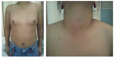
Female 20 years, genetic load for DM and have product of g3 uncomplicated, mother of 23 years. Normal psychomotor development and IQ. Short stature, Without development of secondary sexual characteristics, with f 130/80 primaria. EFTA amenorrhea, c.80 x min. Height: 132cm Weight: 38kg, conjunctive with good color, epicanto, arched palate, Wide neck, short, low hairline, broad chest, breast hypertelorism, absence of secondary sexual characteristics. (Figures 2–4).

Figure 2 Rhythmic heart sounds with aortic expulsivo, and 2 fixed and split noise. Karyotype 46, X, i (Xq).
Pelvic ultrasound, uterus and left ovary small. Ovary Uterus 3.2x1x0.9 left. 1.6x1x1 absent law. TSH 9.25, T4T 8.76, 1.25 FT4, TT3 1.28. FT3 3.46 FSH 97.34. 26.82LH, Estradiol <5, Progesterone <0.03. Prolactin 6.03. Testosterone <0.025 Normal renal ultrasound Bone age 15 years. Levothyroxine and estrogen treatment is started in March 2014.
Early diagnosis is essential: the suspicion is based on clinical signs (physical appearance), but require cariotipo.Una after carrying out genetic diagnosis, detect associated comorbidities. Track for early diagnosis of complications and institute treatment. multidisciplinary monitoring. Full information to parents as soon as possible and the child by his parents, as requested and appropriate content to your age. Short stature, lack of secondary sexual characteristics and infertility condition the lives of patients. It is essential to optimize GH therapy and hormone replacement therapy with estrogen and progestin.8,10
None.
The author declares that there is no conflict of interest.

©2018 Ramírez, et al. This is an open access article distributed under the terms of the, which permits unrestricted use, distribution, and build upon your work non-commercially.