Journal of
eISSN: 2373-4396


Research Article Volume 19 Issue 3
 Anna Chostko, David Sharer, Yuteng Wang, Hisham Qureshi, Aarnav Grover, Annette Kirchgessner
Department of Medical Sciences, Hackensack Meridian School of Medicine, USA
Correspondence: Dr. Annette Kirchgessner, Department of Medical Sciences, Hackensack Meridian School of Medicine, Nutley, New Jersey, 07110, USA, Tel (201)962-5523
Received: June 27, 2026 | Published: July 23, 2026
Citation: Downey M, Chostko A, Sharer D, et al. Tortuous brachiocephalic artery. J Cardiol Curr Res. 2026;19(3):73-76. DOI: 10.15406/jccr.2026.19.00646
Brachiocephalic artery (BCA) variations are rare anatomical anomalies that can increase the risk for perioperative complications by decreasing vascular access and cannulation in vascular and cardiothoracic procedures. This research study examines the occurrence of BCA variations in adult formalin-fixed cadavers dissected as part of a gross anatomy course. This study is among the first to present a high-quality cadaveric dissection of a rare tortuous BCA in an 89-year-old female cadaver with a history of cardiovascular disease. The tortuous BCA measured 1.94 cm in diameter with a 6.1 cm circumference along its tortuous base off the aortic arch. Such manifestations are often asymptomatic but may pose iatrogenic risk across various surgical contexts if unidentified prior to intervention. We consider the importance of preoperative imaging to identify anatomical variations in BCAs to streamline surgical planning.
Keywords: brachiocephalic artery, arterial tortuosity, cadaveric anatomy, aortic arch variants
BCA, brachiocephalic artery; TBA, tortuous BCA; AA, aortic arch; LCC, left common carotid; LSA, left subclavian artery; LVN, left vagus nerve; RCC, right common carotid; RS, right subclavian artery; RVN, right vagus nerve; RVA, right vertebral artery; Tr, trachea; TG, thyroid gland.
The BCA, also known as the innominate artery, is a major artery in the cardiovascular system that carries blood to the right arm, head and neck.1,2 It divides into the right subclavian (RS) and right common carotid (RCC) artery. Variations in its anatomy are particularly important during cervical and mediastinal procedures given its course through the superior mediastinum and anterior to the trachea (Tr).2-5
Anatomy of the aortic arch (AA) and its various branches is established through an embryologic process that involves selective persistence and regression of the pharyngeal arch arteries.6 The BCA arises from the right horn of the aortic sack; its final configuration is determined by normal remodeling of adjacent embryonic vessels. Variations in its course or position may result from abnormal regression or persistence of these surrounding embryonic structures, leading to altered spatial relationships with cervical and mediastinal anatomy.6,7
Anatomical variations of the BCA, including tortuosity and abnormal course, have been described in both clinical and cadaveric contexts and are more frequently observed in elderly individuals.4,5,8 Deviations from normal anatomy may place the artery in closer proximity to the Tr, thyroid gland (TG) and surrounding soft tissue, thus increasing the risk of underrecognized vascular injury during airway management, thyroid surgery, and endovascular interventions.4,9-12 Although many of these variations are asymptomatic and incidentally discovered, their presence can have significant procedural implications.10-13
While prior reports have focused on procedural challenges and clinical complications associated with BCA deviations, fewer studies have characterized these variants through detailed cadaveric examination.3,9,14 Cadaveric findings allow for unique documentation of anatomical relationships that may not be fully appreciated on imaging alone; this can inform surgical awareness and decision-making in addition to mitigating risk.3,8,14
Variations of the BCA were examined during routine dissection of cadavers in the Anatomy Laboratory of Hackensack Meridian School of Medicine. The cadavers donated their bodies to science so that anatomical research could be performed. Following skin and superficial fascia removal, the right sternocleidomastoid (SCM) was reflected to reveal the BCA. The BCA was followed superiorly to where it joined the RCC. At approximately the fourth cervical vertebra, the common carotid artery splits into the external and internal carotid artery.
Sixteen cadavers in the anatomy lab were examined. Five cadavers were male and 11 were female. Each cadaver had its rib cage displaced to allow for observation of thoracic structures, namely the aortic trunk and its associated structures. Each cadaver was subjected to the following processes for the measurement of blood vessels, since it appeared that the tortuous BCA was larger than the normal BCA.
As an indirect measurement of the diameter of a blood vessel, we identified a standard landmark and calculated the value based on the measured circumference.15 To model this, a piece of string was wrapped about the circumference of the BCA. This loop was then brought as close as possible to the juncture formed from the aortic trunk, representing the standard landmark. A pen was used to mark off the string, indicating where this loop closed. The unraveled string, representing the circumference of the BCA was then measured with an accuracy of 0.05 cm.
Using the formula C = πd, where C is the measured circumference, d is the diameter, and π is the geometric constant Pi, the diameter of each BCA was calculated.
Gao et al.16 established a method for the direct measurement of the angle of blood vessels, often performed using image editing software on pictures and measuring angles between lines drawn corresponding to the vessels. The angle between the tortuous BCA and the aortic trunk was measured by pinning 3 points of the arch (one pin at the apex of the tortuous BCA, one pin to the right of the apex pin, and a final pin diagonally superior and to the right of the apex pin), ensuring the arch was positioned in a flat plane. String was then wrapped around each pin to form an angle such that the apex pin served as the angle’s vertex. Next, a protractor was placed at the vertex so that the angle could be recorded.
A third set of measurements was performed. The vessel diameter was measured using a caliper at a consistent position across the cadavers relative to a landmark.17 Again, the landmark used was the branching point for the BCA from the aortic trunk.
During routine cadaveric dissection, a tortuous BCA was found in an 89-year-old female cadaver with a history of cardiovascular disease and pacemaker placement (Figure 1A and 2A). The BCA formed a “buckled” configuration anterior to the tracheal midline and inferior to the thyroid isthmus. As it ascended, the vessel passed posterior to the TG. The RVN maintained its anatomical course but exhibited a closer proximity to the BCA due to the tortuosity observed. The bifurcation of the BCA into the RCC artery and right subclavian artery (RS) appeared in a higher position than typically expected. The artery was measured as 1.94 cm in diameter with a 6.1 cm circumference along the tortuous segment. No additional aortic arch anomalies or evidence of compression of adjacent structures were identified.
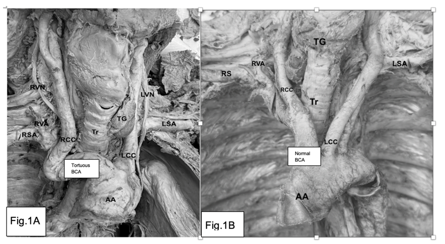
Figure 1 A. Tortuous BCA (TBA). AA, aortic arch; LCC, left common carotid artery; LSA, left subclavian artery; LVN, left vagus nerve; RCC, right common carotid artery; RSA, right subclavian artery; RVA, right vertebral artery; right vagus nerve; TG, thyroid gland; Tr, trachea
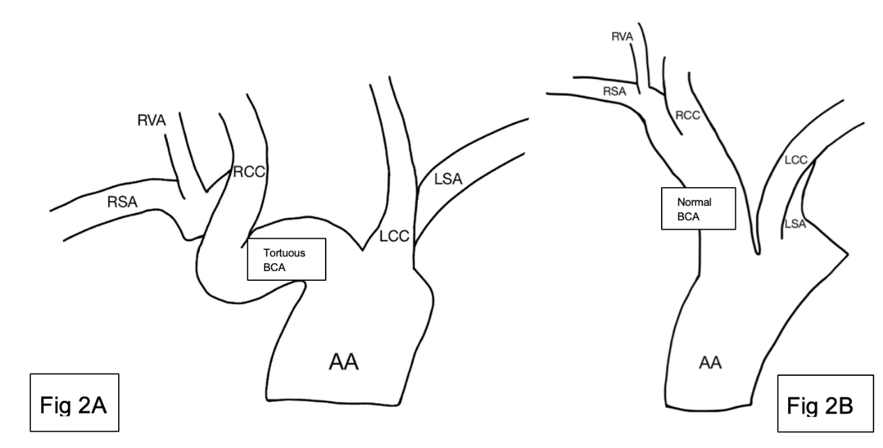
Figure 2 A. Drawing of tortuous BCA (TBA) and surrounding structures. AA, aortic arch; LCC, left common carotid artery; LSA, left subclavian artery; RCC, right common carotid artery; RSA, right subclavian artery; RVA, right vertebral artery; B. Drawing of normal BCA (NBA). AA, aortic arch; LCC, left common carotid artery; LSA, left subclavian artery; NBA, normal brachiocephalic artery; RCC, right common carotid artery; RSA, right subclavian artery; RVA, right vertebral artery.
The tortuous BCA also exhibited an increased circumference and radius and had a significantly increased size compared to the normal BCA. The average circumference of the normal BCA was 5.25 cm (+- .05). The circumference of the tortuous BCA was 6.1 cm (+- .05). Thus, the tortuous BCA was 13.9% larger than the average BCA. The tortuous BCA had the largest circumference among females, measuring at 6.1 cm, with the largest male BCA measured at 6.2 cm.
The tortuous BCA radius was .97 cm, and the average for the remaining normal 15 cadavers was .85 cm. The standard deviation of the average radius was around .06 cm. With the tortuous BCA at .97 cm and the average of all the BCA at .85 cm the z score for the tortuous BCA was 2.14. A P-value of a two tailed test came out to be .046. The angle of the TBCA was 52°. The angles of the other BCAs were not recorded, but they were all observed. There were no other cases of a tortuous BCA among the remaining cadavers.
From the measurement of the BCA using calipers, the average diameter was recorded as 1.496 cm with a standard deviation of 0.140 cm.
When evaluating the dissected cadavers observed over the course of this study, only one elderly female cadaver demonstrated a tortuous BCA. The current understanding is that arterial tortuosity increases with age, high blood pressure and other cardiovascular risk factors, in addition to female sex.18 When considering the cadaver of interest in this research paper, this is in line with established associations, with the female cadaver being of advanced age and having a significant cardiovascular history, including a pacemaker. Such patients may often need interventions that can vary in the degree to which they are invasive, and the presence of a tortuous BCA could pose a perioperative risk.
Salem et al.19 demonstrated that BCA tortuosity added to the technical difficulty of endovascular procedures. Procedural modification or access site conversion was needed to accommodate for this variable, although the authors did identify measurement of a shortened vertebrocarinal distance via X-ray as a predictor for BCA tortuosity. Where the involved blood vessels would still need to typically undergo resection and installation of needed prostheses, the tortuosity can necessitate extra precautions with maintaining cerebral perfusion and even performing anastomoses external to the thoracic cavity.20 Of note here is the consideration of Laplace’s Law. In the present study, a tortuous BCA presented with a larger radius, and by this function, would lead to a proportionately increased tension in the walls of the tortuous BCA relative to the typical presentation. The tortuosity itself can displace the vessels from their normal orientation, bringing them closer to other structures that are then susceptible to iatrogenic injury. Ankle et al. demonstrated that this led to the BCA “riding high”, so much so that it presented as an anterior neck mass.21 This presentation is generally asymptomatic, but this positions the BCA much closer to the trachea, cervical soft tissues, and other vital structures that could be damaged.
Based on the small sample size of this study, the observed frequency of a tortuous BCA within the study sample is approximately 5.8%, although other studies have observed an incidence of 13.9 %.22 Further study is needed to effectively quantify the risk of complications in the context of the incidence in the general population and adjust procedures as needed to account for this. This would allow clinicians to approach procedures with a more complete understanding of a patient's vascular abnormalities, if any, and plan accordingly. Ultimately, this may result in an overall reduced incidence of complications.
Tortuosity of the BCA is an anatomical abnormality with severe cases being minimally documented. This cadaveric study provides insight into the frequency, impact, and implications of the tortuous BCA. The examination included 16 cadavers that had all been dissected, with only 1 having a tortuous BCA with a 52° kink anterior to the trachea and superior to the aortic arch. The observation of a tortuous BCA recorded was around 5.8%. The tortuous BCA had a circumference of 6.1 cm, which was 13.9% larger than the cohort average of 5.25 cm.
The increased circumference and radius of the tortuous BCA have implications for the tension and risk of injury of the vessel. According to Laplace's law, which relates the tension in a cylindrical vessel to the transmural pressure and radius of the vessel, an increase in blood vessel radius directly proportionally increases the tension of the vessel wall. This could predispose the artery to a risk of atherosclerosis. The kink of the tortuous BCA has the potential to disrupt normal blood flow and increase the likelihood of damage happening to the artery. Previous case reports have suggested that brachiocephalic artery tortuosity can lead to fatal complications during tracheotomies. The tortuous BCA increases the complexity of trans radial cardiac catheterization and needs special considerations for imaging before the procedure can be initiated.
While this research study provides valuable morphometric data and clinical implications, the limitations of the study include a small sample size, a lack of complete medical history, and constraints for measuring the arteries. Additionally, the measurements were made ex-vivo using cadavers, inherently being distinct to the vessels found in a living person from the different variables at play. Future research with larger cohorts and more accurate prospective imaging would shine a light on the relationship between a tortuous BCA and its clinical implications.
In conclusion, a tortuous BCA represents an anatomical variant that poses preoperative and clinical risks for patients with the condition. Recognition of the abnormality through MRI and other medical imaging techniques could prevent iatrogenic injury, improving the outcomes of patients who suffer from a tortuous BCA.
We thank Dr. Stanley Terlecky for critically reading the manuscript.
The authors sincerely thank those who donated their bodies to science so that anatomical research could be performed. Results from this research can potentially increase mankind’s overall knowledge, which can then improve patient care.
No conflicts of interest exist.

©2026 Downey, et al. This is an open access article distributed under the terms of the, which permits unrestricted use, distribution, and build upon your work non-commercially.