Journal of
eISSN: 2373-4396


Case Report Volume 18 Issue 4
 Neelam Dahiya,2 Manphool Singhal1
1Department of Radiodiagnosis and Imaging, Postgraduate Institute of Medical Education and Research, India
2Department of Cardiology, Postgraduate Institute of Medical Education and Research, India
Correspondence: Arun Sharma, Department of Radiodiagnosis and Imaging, Postgraduate Institute of Medical Education and Research, Sector-12, Chandigarh 160012, India, Tel 91 9910380907
Received: December 15, 2025 | Published: December 31, 2025
Citation: Sharma A, Dahiya N, Singhal M. Major aorto-pulmonary collateral artery aneurysm in a patient with cyanotic congenital heart disease. J Cardiol Curr Res. 2025;18(4):217-218. DOI: 10.15406/jccr.2025.18.00635
Aneurysms of aortopulmonary collateral arteries are rare and may cause complications including rupture and mass effect on adjacent structures. MDCT helps in clearly depicting the anatomical details of the major aortopulmonary collateral arteries in cases of cyanotic congenital heart disease, thereby helping in choosing optimal management. We, hereby, describe a case of ventricular septal defect with pulmonary atresia with major aortopulmonary collateral arteries acting as source of pulmonary arterial supply in this case with one of the collaterals showing aneurysm.
Keywords: MAPCA aneurysm, ventricular septal defect, pulmonary atresia, cyanotic congenital heart disease, multidetector computed tomography, CT angiography, aortopulmonary collaterals, collateral artery stenosis, aneurysmal dilatation, rupture risk, surgical management
Major aortopulmonary collateral arteries (MAPCAs) are collaterals which supply blood to the pulmonary vasculature when the native pulmonary artery circulation is either atretic or stenotic and is underdeveloped.1,2 Aneurysms of MAPCAs are rare, their early detection is important as they can cause life threatening complications including rupture.3
A 12 year old girl, with a diagnosis of ventricular septal defect with pulmonary atresia, underwent CT angiography for anatomic evaluation of pulmonary arteries, collaterals and the coronary arteries prior to surgical management. She was symptomatic since her childhood with cyanosis and effort intolerance. On examination, clubbing was observed. Oxygen saturation was 70% at room air. CT angiography showed features of large subaortic ventricular septal defect (~18mm) with pulmonary atresia. Confluent small pulmonary arteries were seen with no patent arterial duct. Three large MAPCAs were seen arising from the descending thoracic aorta & the left subclavian artery, two were seen to supply on the left side and one on the right side with areas of stenosis along their course. One of the MAPCAs showed aneurysmal dilatation along its course on the left side. No rupture was seen. No compression was seen on the left bronchus and its branches (Figures 1&2).
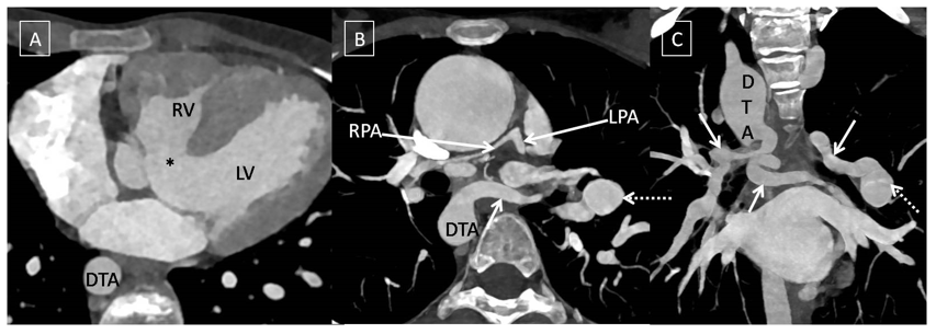
Figure 1 CT angiography images (A-C) showing large subaortic ventricular septal defect (*) with pulmonary atresia and confluent small pulmonary arteries. Major aortopulmonary collateral arteries (solid arrows) were seen arising from descending thoracic aorta (DTA) and the left subclavian artery with stenosis along their course and one of the collateral artery showing aneurysm (dashed arrow).
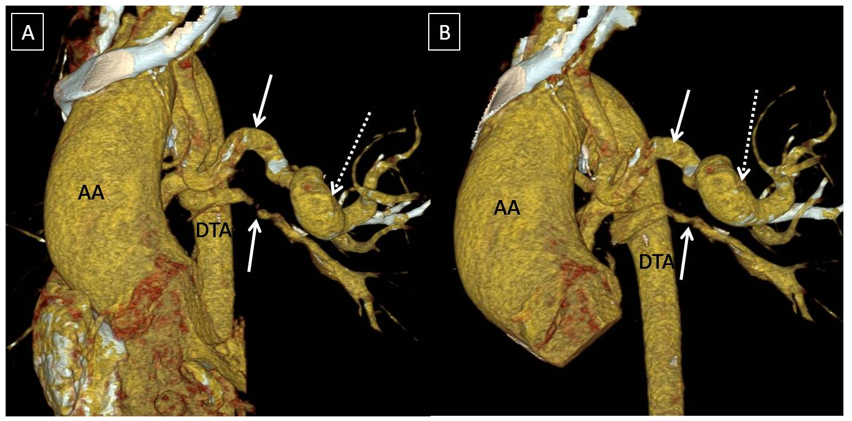
Figure 2 Volume rendered CT images nicely depicting the major aortopulmonary collateral arteries along with the aneurysm.
MAPCAs are aortopulmonary collaterals which commonly arise from aorta and supply the pulmonary vasculature in cases of underdeveloped native pulmonary arteries.1 MAPCAs are frequently seen in cyanotic heart disease with a prevalence of nearly 35% according to an angiographic study by Santos et al.4 MAPCA aneurysms are rare with few case reports in the literature so far.5 They may present with complications including respiratory distress or rupture, or they may exert pressure symptoms on adjacent structures owing to their size.3 In our case, the aneurysm was asymptomatic and was incidentally detected on CT. The CT report should highlight all important aspects of MAPCAs including their number, vessel of origin (aorta, major aorta branches, bronchial artery, and coronaries), O’clock position, course, stenosis, aneurysm, supply etc. for optimal management.6,7
MAPCA aneurysms are rare and may cause life threatening complications including rupture. MDCT evaluation can provide detailed description about MAPCAs which is valuable for choosing optimal management.
None.
Authors declare that there is no conflicts of interest/s to declare.
Manuscript has not been submitted elsewhere or under consideration for publication

©2025 Sharma, et al. This is an open access article distributed under the terms of the, which permits unrestricted use, distribution, and build upon your work non-commercially.