Journal of
eISSN: 2373-4396


Case Report Volume 18 Issue 2

1Division of Cardiovascular Medicine, Hillel Yaffe Medical Center, Israel
2Cardiac Surgery, Rambam Medical Center, Israel
3The Ruth and Bruce Rappaport Faculty of Medicine, Technion, Haifa, Israel
4 Orthopedic Department, Hillel Yaffe Medical Center, Israel
Correspondence: Ariel Roguin MD PhD, Division of Cardiovascular Medicine, Hillel Yaffe Medical Center, Hadera, Israel, Tel 972-4-7744656, Fax +972-4-7744182
Received: July 11, 2025 | Published: July 26, 2025
Citation: Mohsen J, Bolotin G, Behrbalk E, et al. From Backache to Heartbreak - cement embolization from kyphoplasty, resulting in severe tricuspid valve regurgitation and right sided heart failure. J Cardiol Curr Res. 2025;18(2):57-59. DOI: 10.15406/jccr.2025.18.00624
Vertebral body cement augmentation (Kyphoplasty / Vertebroplasty) for treatment of pathological vertebral fracture is a common procedure. Cement leakage to the venous system is a rare complication. We present a case of cement embolization to the right atrium and right ventricle, resulting in tricuspid valve involvement, and right heart failure.
A 59-year-old woman was hospitalized due to dyspnea with pulmonary edema, ascites, severe signs of peripheral edema and liver fibrosis. Echocardiography showed a foreign body attached to the right ventricle, prolapsing into the right atrium and causing severe tricuspid valve regurgitation. CT and CMR showed a foreign body attached to the right ventricle, crossing into the right atrium. She was treated with diuretics and heart failure medications and improved clinically.
Six years prior, she had a history of kyphoplasty due to multiple osteoporotic collapsed vertebrae at the age of 53. Spine CT at the time of cement injection was otherwise normal. In retrospect, a CT done two years later at the age of 55, showed a foreign body in the heart, however at that time she was asymptomatic. Her leg edema developed slowly as well as her liver dysfunction.
The foreign body was surgically removed, and the tricuspid valve was replaced with a biological valve. The foreign body measured almost 10 cm, crossed the tricuspid valve and was embedded within the right ventricular wall. In pathology it resembled bone (appearance on pathology, not histologically confirmed).
In conclusion, intra cardiac cement embolism following percutaneous kyphoplasty is a rare but life-threatening complication. Although often occurring immediately after the procedure, embolisms can also present as a late complication, years after the initial treatment. We present a case of severe tricuspid valve regurgitation caused by the long-term presence of an intra-cardiac foreign body - a cement embolus.
Keywords: cardiomyopathy, complications, heart failure, surgery, tricuspid valv
Percutaneous kyphoplasty and vertebroplasty are minimally invasive surgical techniques that are broadly used to treat osteoporotic vertebral compression fractures and at times also in cases of spinal metastases.1-3 Vertebroplasty involves the injection of bone cement directly into fractured vertebral body. While in kyphoplasty, a balloon is first inserted and inflated into the vertebral body making a space for the cement injection, then the balloon is deflated and removed; after which the cement is injected into the premade space. Both methods contribute to the stability of the vertebra and prevent further fracture collapse and may help in relieving pain.
However, percutaneous cement injection may be associated with multiple complications due to leakage of cement either locally or into the peri-vertebral venous system. The rate of venous cement embolism reportedly ranges from 2.1% to 26.0%.4-7 The cement may rarely flow into the right side of the heart and to the pulmonary vessels.8-11 We herein report a case of late post-kyphoplasty intracardiac cement embolisms that caused severe complications.
A 59 years old female patient, with no known history of cardiac diseases was hospitalized with dyspnea and ascites. She noticed leg edema for a very long period, with worsening in the 4-month prior hospitalization. Dyspnea appeared in the days before and brought her to the hospitalization. Physical examination was positive for significant leg edema, jugular vein congestion, signs of ascites and of pulmonary edema.
Noteworthy, she had severe osteoporosis in part contributed by the chronic steroids use for allergic purpura. Therefore 6 years before the current hospitalization, due to seven vertebral body fracture from T11 to L5, at the age of 53 years old, she was treated by performing two stages kyphoplasty with intravertebral cement injection. First stage injection of cement from L3 to L5 vertebrae, and after 5 days second stage injection from T11 to L2 vertebrae. The two stage procedure was performed to reduce the risk of iatrogenic fat embolization. Percutaneous kyphoplasty was performed via a bilateral transpedicular approach under general anesthesia. Using fluoroscopic guidance between 4.0 to 8.0 mL was injected for each vertebra. No extravasation of bone cement outside of the vertebral body was observed on either anteroposterior or lateral fluoroscopy but some cement leaked into the Anterior External Vertebral Venus System with no leakage into major vessels observed on follow-up radiographs. The patient’s back pain was soon relieved, and she was discharged from the hospital 1 day after surgery.
ECG showed Sinus Tachycardia, and chest X ray showed pulmonary vein congestion with several vertebra showing past cement injection. Some of the cement was visualized as gentle threads parallel to the spine. Another thread was on the heart silhouette. Transthoracic echocardiography showed a foreign body attached to the right atrium and right ventricle near the location of the tricuspid valve, which caused severe tricuspid valve regurgitation (Figure 1 & Movie 1). The patient was taken to the catheterization laboratory and fluoroscopy showed an intra-cardiac foreign body fixed to the heart muscle but moving according to the heart cycle (Figure 2 & Movie 2).
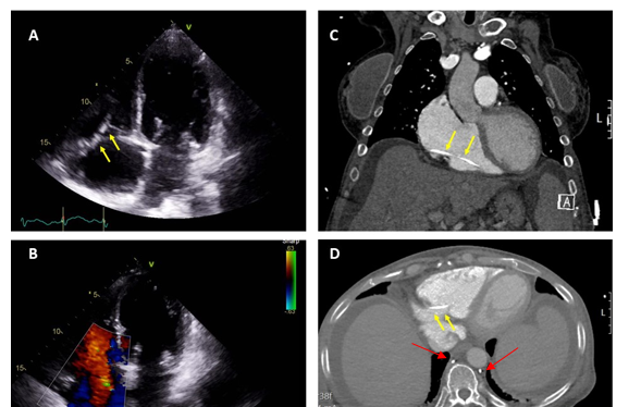
Figure 1 (A) Transthoracic echocardiogram showing the foreign body (arrow). (B) Transthoracic echocardiogram color showing severe tricuspid regurgitation. (C) Cardiac CT showing the foreign body at right atrium and right ventricle (arrow). (D) Chest CT showing the foreign bode inside the right heart (yellow arrow) and from both side of vertebra (red arrow).
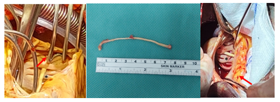
Figure 2 (A) Surgical view of the foreign body (arrow). (B) The foreign body after surgical removal. (C) Close surgical view of the foreign body (arrow).
At this point it was decided to performed some further imaging to characterize the intracardiac finding; Cardiac CT angiography showed mild irregularities in the coronary arteries and a bone density foreign body in the right ventricle. Similar structures were also seen in the paravertebral area on both sides located in the paravertebral veins.
Cardiac MRI showed that the chambers of the heart were enlarged. A strained hyperdense structure about 8 cm long was located in the RV.
During her hospitalization the patient was treated with IV Furosemide with good response, significant weight reduction and improved signs of right side failure. She improved clinically.
Her liver function tested improved significantly, INR was normal, although the albumin was 2.5 mg/dL and did not increase with time in-spite of high protein diet. Repeat Fibroscan of the liver showed that there was liver fibrosis but part of it was due to the congestion caused by the severe TR.
In a revision on previous imaging studies, spine CT at the time of cement injection was otherwise normal. However, in retrospect, a CT done two years after the cement injection, at the age of 55, showed already a foreign body in the heart, however at that time she was asymptomatic. Her leg edema developed slowly as well as her liver dysfunction.
After heart team discussion it was decided to refer the patient for open heart surgery in order to extract the foreign body, and treat the tricuspid valve according to the findings during the operation.
The foreign body was surgically removed, and the tricuspid valve was replaced with a biological valve [Carpentier-Edwards PERIMOUNT Magna EaseVR- size 33]. Transesophageal echocardiography prior to the operation showed a foreign body attached to the right ventricle near the location of the tricuspid valve, which caused severe tricuspid valve regurgitation but not reaching the inferior vena cava. The foreign body which measured almost 10 cm in length and 0.2 cm in diameter crossed the tricuspid valve and was embedded within the right ventricular wall.
Post-operative course was characterized by tissue hypoperfusion and worsening liver dysfunction although both heart ventricles contracted well, the valve functioned properly, and oxygenation was good. On post-operative day 3 she developed DIC with significant acidosis and died due to severe bradycardia.
In patients treated with either kyphoplasty or vertebroplasty for osteoporotic collapsed vertebrae, an unwanted complication may be that the cement injection itself may increase the risk of fractures in neighboring vertebral bodies.1 Additionally, cement leakage from the vertebral body may occur. Cement leakage into the surrounding soft tissues is mostly asymptomatic. Rarely could potentially cause new neurological symptoms if the cement leak presses on the spinal cord or nerves. Leakage into the perivertebral venous system can cause a variety of complications, including rare and life-threatening pulmonary and intracardiac arterial embolisms. Through the paravertebral or epidural veins, the cement leakages into the venous system and further migrate into the right heart and pulmonary arteries. Most cases of cement leakage are considered to be subclinical problems. When occurring immediately after the procedure, the clinical presentation may be abrupt and associated with acute hemodynamic instability.9
However, as in the present case, embolisms can also present as a late complication, even years after the initial treatment. As these embolisms are usually small and do not lead to clinically relevant symptoms, the exact incidence of embolization remains unclear.12
The treatment of choice for symptomatic intracardiac embolism seems to be surgical removal because of the potential complications that can occur.11-13 This is true especially in case of tricuspid valvular involvement, as additional valve repair or replacement may be necessary. We discussed the option of removal using a snaring device but were afraid from break of the foreign body and/or dislodgment and the fact that we may harm the valve apparatus or damage the muscle wall if the cement was inserted deep in the muscle. Another thought was to implant percutaneously the recent Transcatheter bicaval valves system [TricValve] or other options, but opted for a complete removal and perform an established and well recognized treatment. In our case the surgery itself went smoothly, the cement was removed completely, with successful replacement of the valve and good heart contraction.
With the increased usage of cement in the percutaneous treatment of vertebral compression fractures, a higher rate of intracardiac embolisms due to cement leakage is to be expected.
All authors had access to the data and a role in writing the manuscript
None of all the authors

©2025 Mohsen, et al. This is an open access article distributed under the terms of the, which permits unrestricted use, distribution, and build upon your work non-commercially.