Journal of
eISSN: 2373-4396


Research Article Volume 14 Issue 5
Department of Chemistry, University of Antofagasta, Chile
Correspondence: Isabel Pizarro Veas, Laboratory of Bioinorganic and Environmental Analytical Chemistry, Department of Chemistry, Faculty of Basic Sciences. University of Antofagasta, Antofagasta, Chile
Received: August 28, 2021 | Published: October 29, 2021
Citation: Veas IP. Arsenic binding proteins in cardiovascular human tissues. J Cardiol Curr Res. 2021;14(5):137-143. DOI: 10.15406/jccr.2021.14.00530
The intracellular As-protein binding in cytosol and methanol–water extract of the auricle and saphene tissues of as impacted people was evaluated by bidimensional size exclusion FPLC-UV-ICP-MS. The fractionation of cytosol using Superdex, Phenomenex and MonoQ HR 5/5 columns, shows that as is distributed in a wide range of contiguous fractions of each column, being 8, 25, 50 % the percentages of As in the collected fractions, respectively.
In the methanol: Water extracts a similar study than performed with the cytosol using preparative gel chromatography on Sephadex G-75 and Shephadex G-100 columns and the MonoQ HR 5/5 anion protein exchange was carried out. A very low as (<1 % of total As) and protein contain were found in the different fractions of both SEC fractionating series. A similar As–protein association to that found in the cytosol after fractionating with MonoQ HR 5/5 was observed for auricle and saphene. Inorganic and methylated As speciation in the 20 - 26 cytosol fractions obtained within the Phenomenex column was performed by HPLC–ICP–MS using the Hamilton PRP-X100 column. Only As (III) and As (V) were present and the results obtained shows that the As (III)/ as (V) ratio is constant in most cases. Direct evidence of the existence of As–binding peptides in auricle and saphene vein from arsenic impacted human beings has have been obtained which was previously reported by means of novo peptide synthesis.
The danger of as for human health is associated with cancer and non-cancer effects; apart from that its genotoxicity is broadly reported.1 However also a certain essential character had to be assigned to As, in this case, experiments with mammalian model species,2–4 not associated to enzymes. Up to date no metalloprotein of as carrying out some biochemical functionality has been isolated or characterised. The essentiality of as in humans is controversial, being this element able to destroy cell structures, which may benefit or not human health.5,6 Perhaps, the most remarkable threat to human health is cancer, and the most beneficial effect of As2O3 in the treatment of leukaemia is the programmed cellular suicide or apoptosis.7– 9
Among others, two inorganic biochemical paradigms of the behaviour of As must be understood in prokaryotic and eukaryotic systems, as well as mammals, in particular in the human being, who seems to present a special susceptibility for this element. On one hand there is the incorporation and accumulation of as in cells, and on the other, its expulsion from the cells. Both processes are related to essentiality of arsenic, which has yet not been thoroughly demonstrated for humans.6 As(III) is toxic because it is able to form organo-metal bondings with metal-thiol sidegroups in vicinal cysteines of enzymes such as pyruvate dehydrogenase at their activity centres.13 As(V) disrupts oxidative phosphorilation by substituting phosphate in the formation of ATP.14 In some microorganisms arsenical resistance (ars) “operons” involves overproduction of intracellular thiols, which are cytosol proteins that is able to bind As as a first step of a detoxification mechanism.15–18 As binding to various cytosolic proteins and methylation through the methylating agent S–adenosylmethionine (SAM) governed by the ATP as well as arsenite and arsenate methyltransferase are considered competitive mechanisms for As detoxification.19 However, compelling experimental evidence obtained by several laboratories is suggesting that biomethylation, particularly the production of methylated metabolites that contain As(III), is a process that activates as a toxin and a carcinogen.20 Thus, there is evidence that the attachment of inorganic As to proteins and subsequent arsenic extrusion mechanisms by means of cytosol As binding proteins may be the prevalent detoxification mechanism.
Most of the studies with mammalians to investigate the as – protein association have been carried out using “in vivo” or “in vitro” techniques with rabbits (Flemish Giant) or mice. When animals are used for “in vivo” studies, usually increasing doses of As (V) are intraperitoneally injected over certain period. Then the distribution of As(V), As(III) and their methylated metabolites in kidney tissue, liver, lung or spleen are evaluated.19,21–23 Liver cytosol has been the main organ used for these “in vitro” studies because of its important role in detoxification mechanisms.23–27
In this work we have evaluated the intracellular As-protein binding by size exclusion (SE)-Fast Protein Liquid Chromatography (FPLC) with UV and ICP-MS detection in auricle and saphene tissues of the same group of persons in order to understand the effect of long term exposure to high concentrations of as on the cardiovascular system. The As species present in SE relevant fractions have further been determined by Anion Exchange LC-ICP-MS.28–31
Samples
The auricle tissue and saphene vein of six persons operated in the Antofagasta Hospital of coronary thrombosis and presenting high As content in both tissues were taken for this study. The samples were obtained from a population under study, made up of patients who have lived at least five years in the II Region of Chile.32 Sample preparation were made on the bench of the “clean laboratory” inside a laminar flow hood (Labconco, Purifer Class II) using inert devices such as plastic and Titanium knives, agate grinding mortar, and scalpels, scissors and forceps of surgical stainless steel. After titanium clasp and fats residues removal, the tissues were rinsed with deionised water, separated as single samples and stored at –20 ºC. Specific patient’s characteristics can be found in precedent work32 and also in Table 1.
|
FPLC-UV-ICP-MS |
AELC-ICP-MS |
|
Anion Column: MonoQ HR 5/5 |
Anion Column: Hamilton PRP-X100 |
|
Injection volumes: 200mL |
Injection volumes: 100mL |
|
Flow rate: 0.8 mL min-1 |
Flow rate: 1.0mL min-1 |
|
Mobile Phase (gradient): A: 10mM TRIS-HCl (pH= 7.4) B: 250mM ammonium acetate + 10mM TRIS-HCl (pH= 7.4) |
Mobile Phase (isocratic): 10mM Phosphate ammonium (pH = 6) |
|
Time (min) B% Time (min) B% 0 0 10 17 1.5 2 11 18 2 3 12 99 5.5 4 19 100 6.0 10 23 0 |
|
Table 1 Chromatographic conditions for protein separation by FPLC-UV-ICP-MS and As speciation by AELC-ICP-MS
Instrumentation
Before size exclusion and gel fractionating medium pressure liquid chromatography experiments (SE–GF–MPLC), the homogenisation and centrifugation of the tissues were made at 4 ºC in an agate mortar and a Eppendorf centrifuge 5804 R (Germany). These gel chromatography procedures for protein fractionating were made employing tap water thermostated jacket and inert Merck Superformance Universal Glass Cartridge System (50 x 26mm id) (Germany) packed with Sephadex G–75 and G–100 gels, coupled to a Shimadzu LC– 10AS (Japan) chromatography pump via a Pharmacia Fine Chemicals SRV–3 valve and a SA–50 sample applicator (Uppsala, Sweden), and to a Gilson FC 203 fraction collector (France).
A Hydride Generation Atomic Fluorescence Spectrometer (Excalibur, PSA, UK) to determine the total As content was used.
An Inductively Coupled Plasma Mass Spectrometer (ICP–MS) HP 4500 (Yokogawa Analytical Systems, Tokyo, Japan) was used fitted with a Babington glass nebulizer and a Scott double pass spray chamber cooled by a Peltier system, which was employed as an “off line” detector after gel filtration chromatography (GFC) fractionation, and as an “on-line” detector for arsenic speciation by anion exchange liquid chromatography (AELC-ICP-MS) and for arsenic binding protein fractionation by anion protein exchange, namely Fast Protein Liquid Chromatography (FPLC-UV-ICP-MS). Single ion monitoring at m/z 75 was used to collect the data. All signal quantification was performed in the peak area mode. Ge (10µg L-1) was used as an internal standard for ICP-MS. The possible interference of chloride in standards and samples was evaluated.
AELC for inorganic and methylated As speciation was performed with an HPLC system (LDC Division, Riviera Beach, Florida, USA). 100µL of sample were introduced through a 0.45µm nylon syringe filter into the injection valve Rheodyne 9125 (USA). A PRP-X100 analytical and guard anion–exchange columns (Hamilton, Reno, NV, USA) were used. The column effluent was introduced directly into the nebulizer via a 250mm x 0.5mm (id.) polytetrafluoroethylene capillary tube.
The analytical anion – exchange column MonoQ HR 5/5 (50 x 5mm id.) (Pharmacia Biotech. Uppsala, Sweden) FPLC was used for fractionating of cytosol and methanol: water extract.
A gradient HPLC pump Jasco Pu 2089 plus (Tokyo, Japan) and LKB model 2151 UV detector provided with a 10µL flow cell was used for absorbance monitoring during separation and measurements in the tandem FPLC – UV – ICP – MS.
Superdex (300mm x 10mm) and Phenomenex (300mm x 7.8mm) (Pharmacia Biotech. Uppsala, Sweden) analytical gel filtration columns were used to perform protein fractionating in the cytosol, and the above mentioned Sephadex G – 75 and G – 100 to perform protein fractionating after methanol: water extract.
Solvent evaporation of methanol – water extracts and chromatographic fractions were performed in an Univapo100H – Unijet II system (Uniequip, USA). Sonication, when necessary, was performed in a focused ultrasonic bath (Bandelin Sonopuls HD-2200, Fungilab S.A., USA).
Each arsenic species stock solution containing 1,000g L-1 of As, was prepared by dissolving the respective amount of the pure compounds in water. As (III) and As(V) standards were prepared from sodium arsenite and sodium arsenate (Sigma Aldrich, St Quintin, Fallavier, France), dimethylarsinic acid (DMA) and methylarsonic acid (MMA) obtained were obtained from Merck, and arsenobetaine (AsB) and arsenocholine (AsC) from Tri Chemical Laboratory Inc. (Japan). The stock solutions were kept at 4 ºC in the dark. Working solutions were prepared daily and then diluted with water to the final concentration.
The samples were mineralised for total As determination in Teflon reactor vessels (Savillex Corporation 6138 Minneuka, USA) placed in a conventional lab oven and on an aluminium hot-plate.
Deionized water (Milli-Q Ultrapure water systems, Millipore, USA) was used throughout. High-purity nitric and hydrochloric acids were obtained by distillation of the analytical-grade reagent (Merck) in an I.R. distiller (Berghof, BSB-939IR, Germany).
Proteins used for SEC calibration: Blue dextran 2000 (>2.000kDa); alcohol dehydrogenase (150kDa); bovine albumin (66 kDa); carbonic anhydrase (29kDa), cytochrome C (12.4 kDa); aprotinin (6.5kDa), B – 12 vitamin (1,576 Da), Se-cystine (334 Da), Se-methionine (198 Da) and Se-urea (123Da) (Sigma Aldrich, St Quintin, Fallavier, France) and other chemicals were analytical reagent grade or the highest quality obtainable.
Total arsenic determination and extraction for as species.
Details regarding the mineralization of tissues for total As determination and extraction of inorganic and methylated species in water: methanol extracts are given in previous papers.32,33
Cytosol preparation
The fresh tissue was cut into little pieces, minced and homogenised in 2-3mL of Tris solution [10mM Tris, pH 7.4; 0.1mM phenylmethylsulphonyl fluoride (PMSF) and 25mM NaCl] with an agate mortar. The mixture is maintained for 10 min in an ultrasonic focused bath and the homogenised tissue was centrifuged at 30.000g for 15min to remove unbroken cells, and cell debris. The supernatant was further heated at 55 ºC for 15min to precipitate thermally labile proteins and then centrifuged at 30.000 g for 90 min to obtain the supernatant cytosol. The cytosol preparations used were either fresh or stored for a maximum period of one day at - 20ºC. This procedure is based on a modified protocol of G.M. Bogdan et al.26
Procedures for As-protein fractionating by SEC – GFC and anion exchange protein chromatography in the cytosol and methanolic extracts.
The fractionating for cytosol and reconstituted methanolic extracts was carried out following a procedure similar to that reported by Ferrarello et al.34 The general procedure performed is summarised in Figure 1. 200µL of the cytosol were applied to the Superdex peptide analytical column (100 – 7,000 Da). After the void volumes, 30 fractions of 1mL each were collected using 10 mM Tris–HCl [pH=7.4]– 0.1mM PMSF and 25 mM NaCl as mobile phase at 0.5mL min-1.35 This procedure was repeated 5 times collecting the same fractions together up to a final volume of 5 mL each. The pH of the eluent solution chosen was close to the pH of cytosol to avoid structural changes in the protein during SEC elution.
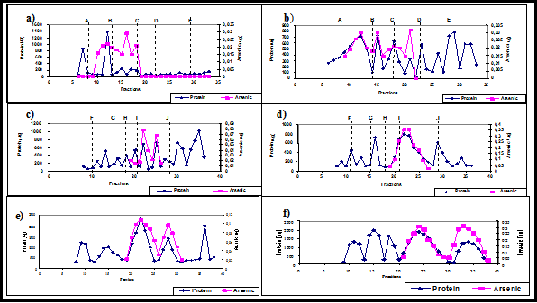
Figure 1 As (µg) and protein (mg) content in the auricle and saphene cytosol of S-1 after fractionating. (a): auricle in Superdex column; (b): saphene in Superdex column; (c) auricle in Phenomenex column; d) saphene in Phenomenex column; (e) auricle in MonoQ HR 5/5 column; (f) saphene in MonoQ HR 5/5 column.
The void volume, (about 30mL), was evaporated up to 1 mL by a N2 vacuum pump at 4ºC. The solution was filtrate through a 0.22µm cellulose filter through vacuum microfiltration, and then it was again size fractionated using the analytical Phenomenex (1,000 – 80,000 Da) column under analogous conditions to those of the Superdex column. The new pooled void volume was evaporated up to 1mL. This volume was fractionated in the MonoQ HR 5/5 anion protein exchange column under analogous chromatographic conditions. The chromatographically separated fractions of the different columns (5mL each), were evaporated up to 3mL and analysed for total protein content by the Bradford´s method36 and for total As by AFS. The fractions having high protein and high As content was further analysed by FPLC-UV-ICP-MS for As – protein binding (200 µL injection volume) and by AELC-ICP-MS for As speciation of As (III), As(V), MMA, DMA, AsB and As C (100µL injection volume). The proteins were measured at 280 nm using an UV detector for the first coupling.
A fractionating procedure similar to that performed for the cytosol was used for the 1:1 methanol: water buffer reconstituted extracts using the Sephadex G-75 (3 – 80 kDa) and G–100 (4–150 kDa) preparative columns, and finally the MonoQ HR 5/5. For the Sephadex columns, 5 mL of sample were introduced on the column head and fractions of 3mL were collected. Only one run was therefore necessary. The dead volume of the G-100 column (about 30mL) was evaporated to 1 mL and introduced after microfiltration into the MonoQ HP 5/5.
TABLA 2 shows the total As concentration of the cardiovascular tissues and some important characteristics of the evaluated impacted persons. Samples S–1 and S-2 were used for the studies performed in the cytosol. Samples S–3, S–4, S–5 and S–6 for the studies performed in the water–methanol batch extracts. Similar results were achieved from additional analysed samples.
Cytosol extracts
Figure 1 (a – f), shows the total As (mg) and protein (mg) profiles of auricle and saphene tissues in the different fractions obtained for S–1 after Superdex, Phenomenex and MonoQ HR 5/5 fractionating. Analogous results were obtained for the auricle and saphene of S–2 sample (data not shown).
A–J dotted vertical lines, show the fraction at which the maximum peak of the proteins set used for calibration appeared. As was distributed within the three columns in a wide range of fractions (about 9 – 22 in the Superdex, 19 – 26 in the Phenomenex and 20 – 34 in the MonoQ HR columns). The similarity for the As and protein profile for both cytosol tissues (especially for saphene) for most of the fractions obtained by the three columns could indicate an As – protein association. The percentages of As in the collected fractions from Superdex, Phenomenex and MonoQ HR 5/5 columns were 8, 25 and 50 %, respectively.
The low resolution of the size exclusion gel fractionating columns does not allow adequate separation of proteins of a small molecular mass difference and therefore neither the identification of the specific protein bound to As. However, it provides information on the protein molecular mass range that binds As. The separation of proteins using the MonoQ HR 5/5, which are 10 µm beaded hydrophilic polystyrene / divinyl benzene resin substituted with quaternary amine groups to yield a strong anion exchanger, is based on the number and nature of ionised side chains of polypeptides. The lack of standards for this column or reference materials makes it difficult to know the molecular mass involved in the different fractions.
In order to evaluate possible As association to the proteins, some representative Superdex, Phenomenex and MonoQ HR 5/5 fractions of both auricle and saphene cytosol tissues were applied to FPLC–UV– ICP–MS.
Methanol: water extracts
1:1 methanol: water mixtures are widely used for As speciation in biological tissues. This extractant mixture was used previously in preparative gel chromatography for arsenosugar detection in algae.36–38 In order to evaluate whether this mixture extracts the As bound to the cytosol proteins in its associated protein form, a similar study than performed above with the cytosol was carried out. A very low content of As (<1% of total As) and protein were found in the different fractions of both SEC / GFC fractionating series. However, a similar association As – protein to that found in the cytosol after MonoQ HR 5/5 fractionating was observed for the auricle and saphene. Figure 2 shows this association for the auricle of sample S-1. The As recovery found within the 25 – 33 fractions (corresponding to the higher MW cutting was 80% of the total arsenic. When FPLC–UV–ICP–MS was applied to the most representative As fractions of MonoQ HR 5/5, chromatograms (not shown) similar to those obtained for the cytosol were obtained. Therefore, we can assume that most of the As content in the methanol: water extract is bound to species that can probably be agglomerated and solubilized in this extract.
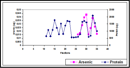
Figure 2 As (µg) and protein (mg) content in 1:1 methanol: water extract after fractionating of auricle S-1 in the MonoQ HR 5/5 column.
As speciation in cytosol and methanol extracts
As speciation within Phenomenex 21–26 fractions of the cytosol from saphene S-1 sample in which an As–protein overlapping occurs was performed to ascertain which As species are attached to the proteins.
AELC–ICP–MS system with the Hamilton PRP–X100 column under conditions given in Table 2, shows the presence of only As (III) and As(V) in all the fractions Figure 3 Similar chromatograms were obtained in the methanol: water fractions of both tissues. The As (III) / As(V) ratio is constant in most of the fraction analysed. A recovery of 90 – 95% [As (III) + As(V)] was found when total As was analysed in each fraction.
|
Total As content in the tissue (µg/g-1) |
Specific patient characteristics |
|||||
|
Sample |
Auricle |
Saphene |
Age of the cardiac infarct |
Working (living) |
As stigmas |
Others |
|
S-1 |
3.9±0.2 |
2.5±0.2 |
37 |
Mine (Calama) |
yes |
Diabetes, dislipidemia |
|
S-2 |
4.5±0.3 |
3.2±0.2 |
44 |
Mine (Chuquicamata) |
yes |
Diabetes |
|
S-3 |
5.4±0.4 |
2.6±0.3 |
45 |
Mine (Calama) |
Yes |
-- |
|
S-4 |
4.8±0.3 |
4.7±0.3 |
50 |
Mine (Antofagasta) |
yes |
-- |
|
S-5 |
4.8±0.4 |
5.2±0.4 |
53 |
Mine(Chuquicamata) |
yes |
Diabetes, anemia |
|
S-6 |
6.1±0.5 |
5.1±0.4 |
65 |
Person from I Region |
no |
-- |
Table 2 Individual information characteristics of the impacted people evaluated and total As concentration (µg g-1), in some of their cardiovascular tissues
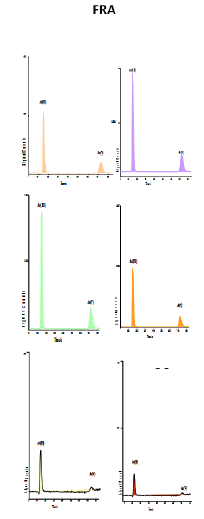
Figure 3 As(III) and As(V) chromatograms from consecutive fractions (20-26) of the Phenomenex column from saphene S-1 cytosol.
It is known that As preferably binds to the sulfhydryl rich cysteine structure, however it must be some instability because the large bound distance (2.25 Å) between As and S. The protonation of sulfhydryl group at the pH of mobile phase (pH = 6) for As speciation could release the As species initially attached to the sulfhydryl rich cysteine structure. The Kd of arsenite for cytosolic proteins was determined by Bogdan et al.26 and the value of 18.4 µM cannot be considered relatively high. On the other hand, the lower pH and characteristics of the stationary phase in the Hamilton PRP X100 column could favoured the protein dissociation.39–42
The toxicity and the mitigation damaging effect may be related with promoting of the folding or degradation of altered proteins or with limiting the synthesis of new proteins that may be altered by arsenite. Another aspect to take into account are detoxification mechanisms of As in the organisms.10,11
The reactivity of As(III), as a soft metal ion which forms strong bonds with functional groups such as thiolates of cysteine and the imidazolium nitrogen of histidine residues, permits to deduce that the understanding of interaction of As ions with proteins is essential in order to understand the mechanisms governing the bioinorganic chemistry related with the biological activity of arsenic. Therefore, independently of some specific protein evolutionary pathways clarified in simple model organisms “phyla”11 common subjects under investigation are: (i) uptake of As(V) by phosphate transporters; (ii) uptake of As(III) in the form of arsenite by aquaglyceroporins; (iii) reduction of As(V) to As(III) by arsenate reductases and finally, (iv) extrusion or elimination of As(III).12,43
The most important findings referring to As binding on proteins may be summarized as follows: i) Arsenic binds a variety of cytosolic proteins and macromolecular constituents of tissues from both, methylating and non methylating (Mamoset monkey) animals;25 ii) As is bound to haemoglobin through vicinal –SH groups in spleen, bone marrow (high haemoglobin content) and in plasma and packed cells. The reduction of arsenate to arsenite is thought to be the major biotransformation mechanism involved;19 iii) As is bound to transferrin, probably at its iron binding sites;19,22 iv) Inorganic As shows a high affinity for fibrous extracellular structures, insoluble structural proteins and the fibrillary tissues with high content in sulfhydryl groups, such as skin and hair.22 This immobilisation could have a protective effect by decreasing the amount of free As (III) capable of interacting with functionally important vicinal sulfhydryl groups of enzymes in the cytosol;22 v) As is bound to 100 kDa, 450 kDa and > 2000 kDa size proteins in liver cytosol in the livers of mammalian animals;14,26 vi) Although most of the arsenite binds to cytosolic macromolecules, a relevant fraction is bound to mitochondrial, lysosomal and microsomal fractions.14,44
Relatively few studies about As binding to proteins in humans have been carried out.27,28 The results obtained with organisms that are assumed to be “good models” of mammals cannot be a priori extrapolated to the human being because these organisms may not realistically reflect the environment of a human body. There is neither a model which correlates the known toxicity of inorganic arsenic with clinical data for symptoms in humans.28 The pharmacokinetic dose–response assessment of a certain trace element cannot be transferred from one species to another without a series of experimental tests.29
The inorganic As content in serum from peritoneal dialysis patients was attached to both, high molecular mass protein (about 80,000 Da) and low molecular mass species (< 1000 Da), whereas arsenobetaine (AsB), although present in the former fraction, was not attached to the proteins.27
There are four arsenic binding proteins of 50, 42, 38.5, and 19.5 kDa. Two of them were tentatively identified as tubulin (50kDa) and actin (42kDa), which are induced by As(III) in human lymphoblastoid cells (28). As(III) binding to proteins is most likely to be an As(III) association to three thiol groups arranged in a specific spatial relationship, as proposed by Rosen.17 Recently, it has been shown by de novo peptide synthesis that As(III)–cysteine interactions stabilise three–helix bundles in aqueous solutions.30 Therefore, the formation of the As(III) complex is likely to cause conformational changes in the proteins to which the metal is bound, an interaction which may be involved in the mechanism of arsenic toxicity.
The high concentration of As associated with copper mining activity and water supply in the Chilean II Region (about 800 mg L-1 in the period 1950-1970, decreasing in the actuality to values around 50µg L-1) is probably realted with occurence of cancer in this region, as well as with non-cancer symptomns such as abnormal pigmentation, acrocyanosis, hyperkeratosis, gangrene of fingers, ischemia of the tongue, diabetes, Raynand´s syndrome, thrombosis, cerebral vascular disease, coronary artery occlusions and other cardiovascular diseases (CVD).1,31,45,46
This work can be considered as a first step in investigation on As bioaccumulation and the biochemical response of cardiovascular tissues proceeding from individuals chronically impacted by inorganic As. When the source for human As uptake is drinking water, As is basically present as As(V) whilst As (III) is the predominant species when air is the source of pollution. Chronically exposed persons often present cancer, beside other toxic effects such as As stigmas on skin and an elevated risk to suffer heart attacks, which always affects the quality of life and may possibly lead to prematurely death.
At the current state it could not be finally concluded to which specific protein As is associated. However, the following findings may help to clarify some aspects of As metabolism by the human body.
For a deeper insight in As – protein association as part of the metabolism pathways additional studies are evidently necessary. Ongoing work is focussed on this aspect.
None.
The rest of the authors declare do not have conflicts of interest.
None.

©2021 Veas. This is an open access article distributed under the terms of the, which permits unrestricted use, distribution, and build upon your work non-commercially.